
Review · Volume 17, Issue 1 · pp. 121–137
Deep Learning Approaches for Accurate Diagnosis and Prognosis of Skin Cancer
Amit Anand1, Aparna Inamdar2, Bannimath Gurupadayya2, Kushala Reddy3, Mansi Sharma4, Mohit Sharma5, Arindam Kolay6, Dipayan Rath7, Romi Kumari Thakur8, Himanshu Sharma9
- 1Department of Pharmacognosy, JSS College of Pharmacy, JSS Academy of Higher Education and Research, Mysuru, Karnataka, INDIA.
- 2Department of Pharmaceutical Chemistry, JSS College of Pharmacy, JSS Academy of Higher Education and Research, Mysuru, Karnataka, INDIA.
- 3Department of Pharmaceutical Chemistry, BGS Global Institute of Pharmaceutical Sciences, Kengeri, Bangalore, Karnataka, INDIA.
- 4Department of Pharmacology, Teerthanker Mahaveer College of Pharmacy, Teerthanker Mahaveer University, Moradabad, Uttar Pradesh, INDIA.
- 5Department of Pharmaceutics, Nims Institute of Pharmacy, Nims University Rajasthan, Jaipur, Rajasthan, INDIA.
- 6S. K Patel College of Pharmaceutical Education and Research, Ganpat University, Kherva, Gujarat, INDIA.
- 7Department of Pharmacology, IQ City Institute of Pharmaceutical Sciences, Durgapur, West Bengal, INDIA.
- 8Department of Pharmacy Practice, Nims Institute of Pharmacy, Nims University Rajasthan, Jaipur, INDIA.
- 9Department of Pharmacology, Nims Institute of Pharmacy, Nims University Rajasthan, Jaipur, INDIA.
Abstract
Skin cancer remains one of the most prevalent malignancies worldwide, and delayed diagnosis often leads to poor clinical outcomes. Recent advances in Artificial Intelligence (AI), particularly deep learning and computer vision, have revolutionized dermatological imaging analysis by enabling rapid and accurate detection of skin lesions. AI-assisted systems have emerged as valuable tools for improving diagnostic precision, prognostic evaluation, and accessibility to dermatological care. This review aims to summarize current applications of AI in the diagnosis and prognosis of skin cancer, highlighting its role in lesion classification, clinical decision-making, teledermatology, and medical education. The review also discusses existing limitations and future perspectives for safe clinical integration. A narrative review of published literature on AI-based approaches for skin cancer management was conducted. Studies focusing on machine learning, deep learning, dermatoscopic image analysis, prognostic prediction models, and telemedicine applications were evaluated. Key areas assessed included diagnostic accuracy, lesion classification, patient monitoring, and ethical or regulatory considerations. AI systems demonstrated high diagnostic performance in differentiating malignant from benign skin lesions and accurately identifying melanoma, basal cell carcinoma, and squamous cell carcinoma. These technologies supported early detection, personalized treatment planning, and prognostic assessment using clinical and imaging parameters. AI-enabled telemedicine platforms improved access to dermatological expertise in underserved regions, while annotated datasets enhanced dermatology training and education. Continuous lesion monitoring reduced diagnostic variability and improved follow-up assessment. However, concerns regarding data privacy, algorithm transparency, dataset bias, and regulatory compliance remain significant challenges. AI-based technologies have substantially advanced skin cancer diagnosis and prognosis by enhancing early detection, diagnostic accuracy, and individualized patient care. Despite existing ethical and validation challenges, continued development and rigorous clinical evaluation of AI tools may significantly improve dermatological practice and patient outcomes in the future.
Keywords: Artificial Intelligence; Basal Cell Carcinoma; Dermatology; Personalized Treatment; Skin Cancer; Telemedicine
FULL TEXT
INTRODUCTION
One of the most prevalent diseases in the world, cancer arises from the rapid proliferation and division of abnormal cells, which in turn destroy normal body cells, causing damage to organs, tissues, and other organs as well as impairment of normal function and eventual cell death. Malignant carcinoma, also referred to as metastatic cancer, spreads from one organ to another, while benign cancer is completely localized and specific to the affected organ (Jones et al., 2022). Cancer and its various forms are divided differently based on their origin in the human body. The different types may be categorized as melanoma, a type of skin cancer, as well as other types, including endometrial, kidney, leukemia, liver, lung, non-Hodgkin lymphoma, pancreatic, thyroid, bladder, breast, colon, and rectal cancers (Salinas et al., 2024). As we all know, skin is the largest organ of the human body, and its cancer leads to uncontrollable and abnormal cell proliferation in the epidermis and uppermost layer of skin. Hence, it should be diagnosed and treated at the earliest stages. For instance, a 2024 Stanford-led meta-analysis of 12 studies with 67,000 assessments revealed that AI-diagnosis enhanced both sensitivity and specificity, particularly for general practitioners and medical students. Nonetheless, this performance was obtained in a selected dataset under specific testing conditions, and may not reflect real-world variability, data imbalance, or potential algorithmic bias. Further large-scale, multicenter studies with diverse populations are required before such performance metrics can be considered universally applicable (Papachristou et al., 2024). The awareness and early diagnosis of skin cancer is critical for the diagnosis of melanoma. Physicians may identify skin cancer. Patients and families notice over 75% of non-melanoma skin malignancies for the first time. Recognizing the early warning symptoms of skin tumors, along with performing frequent evaluations of the skin, aids in the early identification, especially when the patient's condition is not worsened and can be treated at the earliest before it spreads to other organs of the body. Self-skin inspections must be carried out in an illuminated room with a full-length mirror (Menzies et al., 2023). It is beneficial to have another individual examine the head and back part of the body. Non-melanoma cancer of the skin most commonly originates in locations that are regularly subjected to sun radiation in persons with lighter skin that has less melanin content in the body. Artificial Intelligence (AI) is gaining more importance in the analysis of skin cancer, especially in the initial identification and categorization of lesions of the skin. Dermatologists can benefit from AI-powered systems that provide accurate and timely assessments, thereby decreasing human errors and increasing patient outcomes (Vasey et al., 2022). Algorithms based on AI can analyze skin images, such as dermoscopic and diagnostic photos, to identify possibly malignant tumors at an early stage, leading to enhanced therapy and an improved prognosis. AI can properly examine skin lesion photos, looking for traits like inequalities, border variability, color fluctuation, and other signs of probable carcinoma. AI systems examine skin lesions objectively, eliminating the possibility of individual error in diagnosis (Farina et al., 2022). AI can help oncologists make better-informed judgments by offering an additional perspective or preliminary evaluation. AI may be utilized in large-scale operations evaluation programs to efficiently and quickly examine and assess a huge number of skin photos. Artificial intelligence can help telemedicine by allowing remote dermatological consultations and evaluations (Brancaccio et al., 2024).
MATERIALS AND METHODS
The review was conducted using the databases Medline, Embase, ScienceDirect, and Scopus. The search strategy employed the keywords “artificial intelligence,” “skin cancer,” “basal cell carcinoma,” “dermatology,” “telemedicine,” and “personalized treatment.” Additional relevant information was obtained from textbooks, review articles, and other scholarly documents. The study selection process was carried out in accordance with the PRISMA (Preferred Reporting Items for Systematic Reviews and Meta-Analyses) 2020 guidelines, and a corresponding PRISMA flow diagram illustrating the identification, screening, eligibility, and inclusion of studies is presented in Figure 1.
Skin Cancer Basics
Types of skin cancer
Largest organ of the body is the skin in humans, and it regulates body temperature and protects the body from harm and illness (Muncey-Buckley et al., 2025). This organ consists of three basic layers: the epidermis, the outer layer; the dermis, the inner layer; and the hypodermis, a thick layer of fat situated in the skin (Felmingham et al., 2023). The tumors of the skin should be treated at the earliest, since they affect skin dysfunction, followed by impairment of its associated body functions due to the uncontrollable and abnormal cell development in the epidermis and uppermost skin (Jinnai et al., 2020). Melanomas are classified primarily as four types like basal cell carcinoma, squamous cell carcinoma, melanin carcinoma, and Merkel cell carcinoma. Basal cells are globular cells that line the bottom epidermis. These cells are responsible for approximately 80% of skin cancers. Although it can arise at any place on the skin, basal cell carcinoma most commonly presents on the top of the head and neck. The major etiological factors, such as sunlight exposure and radiation therapy, are administered to children (Melarkode et al., 2023). SCC is the second-most identified, as it accounts for approximately 20% of all skin cancers, as the epidermis consists of flat and scaly morphology cells (Hamamoto et al., 2020). SCC can be seen in various parts of the skin, as it often occurs due to sun exposure. The major etiological cause for SCC could be burned and injured skin due to continuous chemical and X-ray radiation exposure, especially, lips wherein a wound has existed for a longer period and skin surrounding the oral cavity, the anus, and the genital area skin are all common sites for squamous cell carcinoma (Han et al., 2022). Approximately 2-5% of SCC instances may spread to other body parts (Djavid et al., 2021). The rarest form of skin cancer is Merkel-cell tumor, which spreads extremely and very rapidly. It is generally observed in the hair follicles and the cells, leading to hormone balancing in the body, and MCC is also known as neuroendocrine skin cancer (Wen et al., 2024). Melanoma is a melanin carcinoma, where melanocytes, generally known as sporadic cells, are found in the intersection of dermis and epidermis. Melanin cells function normally in the body by producing the melanin pigment, essential for skin color and different shadings (Bera et al., 2019). Melanoma, a severe skin cancer, develops in melanocytes and accounts for 1% of skin malignancies. Different stages of melanoma and its progression.
Stage I melanoma is confined to the epidermis, with a thickness of less than 1 mm and no ulceration. At this stage, malignant melanocytes are localized, and there is no involvement of lymph nodes or deeper tissues. Prognosis is generally favorable when detected early, and surgical excision is often curative (Bera et al., 2019). Stage II disease remains localized to the skin but exhibits increased tumor thickness (1-4 mm) and, in some cases, ulceration (Qurban and Cassidy, 2024). This stage carries the poorest prognosis and typically requires systemic treatment modalities, including targeted therapy or immunotherapy (Das et al., 2021).
Causes and risk factors
A malignant tumor occurs when cells begin to multiply and grow uncontrollably. One established cause of the emergence of skin cancer is exposure to sunlight, both natural and artificial (Marsden et al., 2024). The DNA in skin cells can be significantly damaged by Ultraviolet (UV) rays that emanate from sunlight and tanning booths. These damaged cells have the potential to grow into cancer (Shaumya and Kumar, 2025). Despite dangerous Ultraviolet (UV) rays can occur many years before the detection of cancer, it can also occur soon before skin cancer. (For example, an individual who often tans in their early adolescence may get skin cancer several decades or possibly two later. Skin malignancies that form on body parts that are not exposed to the sun, on the other hand, cannot be ascribed to UV radiation (Eskandar, 2023). Cancer of the skin is more probable to impact those who have a lower level of immunity, such as recipients of organ transplants or people who have certain medical conditions. Exposure to particular chemicals, such as arsenic, can upsurge the probable risk of skin cancer (Usmani et al., 2023). Previous cancer radiation therapy increases the risk of skin cancer in the treated area. Men are more susceptible than women to developing skin cancer (Vera et al., 2022). The risk of skin cancer increases with age, with older persons being more sensitive. Spending prolonged amounts in sunlight, especially inadequate precautions like sunblock and appropriate garments, increases the possibility of acquiring skin cancer. A family history of skin cancer may indicate an inherent sensitivity to the condition, possessing a lot of malformations or unusual moles can lead to melanoma, the most severe kind of skin cancer (Karimzadhagh et al., 2026).
Clinical evaluation and staging
In melanoma, clinical assessment and staging are critical for assessing disease extent, guiding treatment options, and forecasting patient outcomes (Alum, 2025). Melanoma staging is frequently performed using the methodology employed by the American Joint Committee on Cancer (AJCC) staging approach. The AJCC 8th edition is the most recent as of my final knowledge update in 2022. It should be kept in mind that the staging system may have been updated or revised after that date (Mar and Soyer, 2018).
Current Diagnostic methods for skin cancer
Once melanoma has been diagnosed, various tests such as lymphatic node visualization and sentinel lymphatic system biopsies, computed tomography, positron emission tomography scan, a magnetic resonance imaging with gadolinium as ultrasound exam, and blood test studies may be conducted to ascertain whether melanoma cells have travelled within the skin or to other parts of the body (Kang et al., 2023). There are three possible methods by which cancer might spread throughout the body. The cancer could spread to other body parts from its original place. The stage of melanoma is determined by the thickness of the tumour, whether the cancer has spread to the lymphatic system or other parts of the body, and other various factors. A representation of some common AI-based diagnostic methods for skin cancer is illustrated in Table 1.
| Methods | Description | References |
|---|---|---|
| Dermoscopic image analysis | Analyzing images captured using dermoscopy, a technique that uses a handheld device with a magnifying lens and light. | (Connor et al., 2025) |
| Convolutional Neural Networks (CNNs) | Advanced deep learning models which perform best at image recognition, are utilized for image-based classification of skin lesions. | (Nahm et al., 2025) |
| Computer-aided diagnosis systems | Systems that assist dermatologists in analyzing skin lesions by providing additional information and suggestions. | (Wei et al., 2024) |
| Automated mole mapping | Using AI to map and monitor changes in moles over time, helping in the early detection of potential skin cancer. | (Tschandl et al., 2020) |
| Texture analysis | Analysing the texture patterns of skin lesions to identify irregularities associated with cancerous growth. | (Tschandl et al., 2020) |
| Clinical data integration | Combining AI analysis with clinical data, including patient history, to enhance the precision of skin cancer diagnosis. | (Chuchu et al., 1996) |
| Mobile Apps for skin lesion monitoring | AI-powered apps that allow users to monitor and track changes in their skin lesions using smartphone cameras. | (Ferrante di Ruffano et al., 1996) |
| Machine learning models | Utilizing various machine learning algorithms to analyze data, including features such as colour, shape, and size. | (Janda et al., 2022) |
| Artificial intelligence in pathology | AI applications in analyzing pathology slides to aid in the diagnosis of skin cancer at the cellular level. | (Kalidindi, 2024) |
| Ensemble learning approaches | Combining estimates from various AI models to enhance overall diagnostic accuracy and reduce false positives/negatives. | (Zhang et al., 2022) |
FUNDAMENTALS OF ARTIFICIAL INTELLIGENCE IN DERMATOLOGY
Machine learning and deep learning
AI has created a great revolution in the field of medicine, especially in the oncology sector, which has a foundation of integrated computer systems (Harrer et al., 2019). AI and its several other models and their algorithms give us various directions for prognosis, diagnosis, and treatment therapy for skin cancer (Longoni et al., 2019). This AI also provides doctors with assistance in understanding the stages of melanoma and predicting the future growth of melanoma by considering the present parameters, patient health conditions, and test results (Mashraqi and Allehyani, 2022). AI has reached a point where it can successfully detect melanoma and its types, stages of melanoma in patients, and treatment options available for the patient to cure skin cancer with the assistance of machine learning and deep learning algorithms. Normalizing the network inputs to ranges between 0 and 1 increases the model's accuracy (Wang et al., 2022). The outputs are considered standard outputs for classification, they yield fractional values in the range of 0 and 1. It works by using Bayes' theorem to predict skin cancer by looking at dermatological pictures with a very high precision rate. Bayes' theorem can be expressed as:
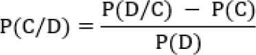
Here;
P(C): probability of developing skin cancer (prior probability).
P(D): Probability of observing the given dermatological features.
P(C∣D): Probability of having skin cancer given the observed features (posterior probability).
P(D∣C): Probability of observing the features given that the person has skin cancer.
P(-C): Probability of not having skin cancer.
This model has an accuracy of around 70.15% along with its assistance to a physician in the analysis and accurate treatment of skin cancer by detecting a particular part of skin disease it mainly works by probability classification model where each output is added into each classifier and then a probability posterior distribution is taken as the outputting (Janda et al., 2020). Overall, three model parameters were identified, namely, the proliferation rate of melanoma cells, the diffusivity of melanoma cells, and the rate of degradation of melanoma cells in skin cancer patients. All three models’ particular algorithms collect information from the 2D circular barrier assay and the 3D invasion assay. This primarily works using a primary classification of problems and extends towards fit-regression problems (Kakish et al., 2025). After a liver transplant, the long-term risk of the development of non-melanoma skin cancer could be detected intuitively by utilizing decision tree algorithms by monitoring the peri-transplant period carefully. Various risk factors are associated with this algorithm, or kidney failure, diabetes, obesity, etc. This AI model has specificity, sensitivity, and accuracy greater than 90%. It is one of the AI algorithms, referred to as the KNN model. Proximity and distance as a matrix are mainly used to classify the data points in a supervised parametric classification algorithm (Górecki et al., 2025). The KNN model is used to detect skin cancer, as well as being able to visualize the tumor correctly. This model provides and quantifies information about all types of skin lesions, without the need to perform biopsies of the skin. There is one hybrid classifier model that is built mainly based on KNN classifiers and predicts the melanoma types mainly based on two methods, those are threshold-based segmentation and the ABCD extraction feature. It uses a firefly optimization, which is coupled with KNN to identify the type of skin cancer computationally, along with an accuracy of 93.33% and sensitivity of 88.89%, along with 100% specificity. This is one of the clustering methods that is usually grouped under an unsupervised learning method, where it employs fuzzy logic with the algorithm of K-means clustering. This model is used over an already processed clinical image of the skin to detect the affected region (Zhang et al., 2026). This algorithm mainly groups the objects to ensure that there is the least variance in each group to make this classifier system a high-featured segmented image. In the initialized class center each pixel of each image is assigned randomly and the centers are recomputed based on every data point inserted and the process is continued to repeat until all the data points have been correctly assigned in the clusters. Once these formed, these clusters could be further subjected and processed to detect the melanoma cancer cells with an accuracy of 90%. This AI model is an extended model of decision trees. This mainly works using a proposed method of the training set that further generates multiple sub-training sets and these works utilize computing the Gini index for every sub-training set followed by the population of the model with decision values. The decision values from every individual are grouped to create a model that classifies the test samples utilizing voting, the class receiving most votes is picked finally in the end step and the outputs are produced. Skin cancer could be detected by describing matrix elements of Mueller by random forest algorithm (Kann et al., 2021). This random forest compared with the decision tree provides enhanced and highly accurate results of 93% along with a reduction of variance. SVM Models predict classifies and extrapolate the data by analyzing data. It utilizes ABCD features for characterizing and extracting the features of legions such as color size shape from the clinically provided pictures. Once these features are selected it classifies the lesions as, keratosis of seborrheic tissue melanoma, erythematosus of lupus melanoma, along with non-melanoma and melanoma skin lesions. But this process mainly having 6 steps or phases those are acquiring the image, preprocessing, extracting and segmenting the features, classification the image, collecting the results, and analyzing them with 98% sensitivity, 95% specificity, and 90% accuracy data analyzing and prediction of carcinoma of skin, machine learning models where it combines the predictions obtained from two or more AI models. The constituent models are those that are the ensemble members of this model, and these ensemble members are grouped in a category and generate an output of a prediction of a particular type of melanoma in a skin cancer-diagnosed patient (Riaz et al., 2024). It utilizes a neural network fuser for grouping of ensemble members and it gives a very good result as compared to the individual classifier models of AI. Table 2 illustrates the different machine learning and AI-based models used in detecting the different types of skin cancer.
| Sl. No. | Type of skin cancer | Machine learning AI model | Description of AI model | Limitations | Reference |
|---|---|---|---|---|---|
| 1. | Non-melanoma | AI neural network AI model | Dataset obtained from the National Health Interview Survey by taking 12 neurons from each layer and further normalizing them to fall between 0 and 1. | This model does not consist of exposure to UV-radiations and does not consider family history during predictions. | (Rivera et al., 2020) |
| 2. | Segmentation and detection of skin disease | Naïve Bayes AI model | It uses a dynamic algorithm, graph cut, for skin lesion and its segmentation by a Naïve Bayes AI model from ISIC 2017. | It cannot be able to differentiate between a few colors. | (Gomolin et al., 2020) |
| 3. | Non-melanoma | Decision tree AI model | COX Regression analysis by decision tree analysis model from the Oregon Procurement Transplant Network 2016. | This model building and its further validation are not from independent cohort charts. | (Cabello et al., 2025) |
| 4. | Skin lesions | K-nearest neighbor AI model | It predicts the type of skin cancer based on threshold segmentation. | Segmentation of an image and its preprocessing is mainly dependent on threshold segmentation values. | (Cabello et al., 2025) |
| 5. | Melanoma | Random forest AI model | Segmentation of skin, followed by skin extraction and detection of lesions as per ISIC. | This classification could be regenerated with a very high accuracy using a simple vector machine. | (C. Zhang et al., 2023) |
| 6. | Melanoma | Simple vector machine AI model | Works by extracting the features of skin such as color texture shape of lesions, as per guidelines of University of Medical Centre Groningen Database. | This model cannot detect the images of the kin with the hair or uncropped images. | (Aravazhi et al., 2025) |
| 7. | Multiple types of skin cancer | Ensemble learning AI model | Human against machine ISIC 2019, by utilizing five deep learning models. | This is a highly imbalanced data set as it leads to improper results during the validation of the model. | (Abdul Rasool Hassan et al., 2025) |
| 8. | Melanoma | A restricted Deep Boltzmann machine AI model | It predicts somatically mutated genes that usually undergo Malignant transformations and gives us results of juggling and mutation profiles of each gene. | The sample size is very short and all the data is presented at a genetic level. | (Troyanskaya et al., 2020) |
| 9. | Detection of skin cancer | Deep auto encoders AI model | This is mainly based on a dataset consisting of auto encoding model followed by reconstruction of spiking networks leading to enhanced results. | This AI model unnecessarily extracts various irrelevant features. | (Bekeschus et al., 2025) |
| 10. | Melanoma | Recurrent neural network AI model | It utilizes modified deep-learning algorithms and optimizing them from RNN's. | This model is completely dependent on segmentation parameters and its classification. | (Liu et al., 2020) |
| 11. | Melanoma, Keratosis of the skin | Deep convolutional neural network AI model | It detects the type of skin cancer utilizing ECOC SVM and CNN deep Images and is further cropped to reduce the noise. | This model does not obey the Asymmetry, Border, Color, and Diameter (ABCD) rule. | (Liu et al., 2020) |
| 12. | Melanoma | Extreme learning machine | Once the preprocessing is completed, the Otsu Method is utilized for segmenting the skin and extracting lesions, followed by the detection. | Highly time-consuming and very intensive AI model. | (Liu et al., 2020) |
Model training and evaluation
This may involve adjusting the learning rate, adding regularization, or changing the model's depth. Assess the last trained model on the test set to estimate its actual performance in real-world scenarios. This gives an unbiased accuracy of the model on unseen new data (Aamir et al., 2024). Understand the model's decisions by employing interpretability techniques. Once the model is trained and evaluated successfully, it can be applied for real-world use. Integration into a healthcare system or a mobile application for skin cancer screening is an example of deployment. This is crucial to involve medical professionals in the model development process to ensure the accuracy and safety of the AI system in diagnosing skin cancer (Miller and Brown, 2018).
DATA SETS AND DATA SOURCES
Publicly available skin cancer datasets
Several publicly available datasets for skin cancer research were accessible. However, the availability of datasets may change over time, and new datasets may become available. Table 3 represents the datasets that were known to be publicly accessible for skin cancer research.
| Available datasets | Description | References |
|---|---|---|
| ISIC (International Skin Imaging Collaboration) Archive | ISIC offers an extensive library of skin images, including dermoscopic images, clinical images, and others. It’s a great tool for creating and validating machine-learning algorithms for the diagnosis of skin cancer. | (Saleem et al., 2025) |
| PH2 | The PH2 Dataset includes dermoscopic images of common pigmented skin lesions. It is useful for research in the automatic diagnosis of melanoma. | (Wang et al., 2025) |
| HAM10000 | This dataset includes over 10,000 labeled images of skin lesions, covering various skin conditions, including melanoma. It is available on Kaggle and is suitable for machine learning research. | (Susanto et al., 2025) |
| Dermo-fit image library | Dermo-fit is a dataset containing standardized images of various skin lesions. | (Kolla and Parikh, 2024) |
| SONIC skin dataset | It can be used for research in computer vision and machine learning for skin disease diagnosis. | (Krishnan et al., 2023) |
AI ALGORITHMS FOR SKIN CANCER DIAGNOSIS
Image classification
Study suggested that a deep learning algorithm on a combination skin dataset of 129,450 clinical and dermoscopic pictures representing 2032 distinct skin lesion disorders produced the most significant advancements in this field. They evaluated how well a deep learning technique performed in classifying and differentiating between benign seborrheic keratoses and carcinomas, as well as between benign nevi and melanomas, using 21 board-certified dermatologists (Hartmann et al., 2024). To classify skin cancer, AI performance was shown to be comparable to that of dermatologists. In the study presented skin lesion categorization and diagnosis are achieved using three primary modalities clinical pictures, dermoscopic images, and histopathological images (Shah et al., 2019).
Image segmentation
Morphological analysis and adaptive thresholding are used to implement segmentation. The threshold for adaptable images is provided by the equations:

Where;
Tx = ½ {miny (maxx f (x, y)} - {max f (x,y)}+ {maxx f (x,y)y}
Ty = ½ {minx (maxy f (x,y)} - { maxy f (x,y)x} + {max f (x,y)}x
Here, x and y represent means in column x and row y, respectively. This method provides a way to extract the most important characteristics connected to a well-defined object within the picture frame.
Ensemble models
The goal of creating ensemble models with diverse learners is to use CNNs' capacity for pattern extraction to address the complexity of multiclass issues and enhance multiclass problem generalization using ensemble systems (Hunter et al., 2022). Through the utilization of individual learners' variability and increased generalization of the learning system, ensemble techniques combine their outputs to improve accuracy. Because of bias and variation, machine learning models are constrained by their hypothetical spaces. To get over a single learner's potential limitations in capturing the distribution (variance) of data, ensemble approaches combine the decisions made by individual learners. As a result, combining the knowledge of several varied learners to make a choice may increase robustness while lowering bias and variation. By aggregating the basic learners, ensemble learning uses a variety of strategies to produce a robust and accurate composite model. Voting, averaging, cascading, and stacking are possible combinations of techniques. The averaging technique comprises both weighted and averaged averaging, whereas majority and weighted majority voting include voting strategies (Maatouk and Lucero‐Prisno III, 2025).
Explainable AI in skin cancer diagnosis
Deep neural network in transparency is frequently addressed with Explainable Artificial Intelligence (XAI). These approaches in dermatology typically provide pictures that resemble the image that is being highlighted. certain image zones that are key for the classification. The application of XAI, however, subjected to an ongoing study (Maru et al., 2024).
PERFORMANCE EVALUATION
Metrics for model evaluation
The suitability of various model components may be determined by utilizing various graphical methods that assess a fitted model. Biomedical research and clinical trial applications, model evaluation often goes beyond simple accuracy metrics (Alzaareer et al., 2024). In this context, the Nonlinear Mixed-Effects Modeling (NLMEM) technique is particularly valuable because it can account for both fixed effects (population-level parameters) and random effects (individual variability) (Moglia et al., 2025). NLMEM enables the modeling of longitudinal or repeated-measures data, which is common in clinical AI validation where multiple measurements are taken from each patient over time. This technique assumes that the model’s structural component describes the underlying relationship between predictors and outcomes, while the random-effects component captures deviations due to inter-individual and intra-individual variability (Martínez-Vargas et al., 2025). Residual error models are also incorporated to handle unexplained noise in the data. By integrating covariates such as age, lesion type, or imaging modality, NLMEM allows for more precise interpretation of AI system performance across heterogeneous populations (Saifan et al., 2025). Evaluation of an NLMEM fit involves both numerical and graphical diagnostics. Goodness-of-fit plots, Visual Predictive Checks (VPC), and Normalized Prediction Distribution Errors (NPDE) help identify model misspecification (Akazawa and Hashimoto, 2020). Importantly, NLMEM supports external validation by simulating data under varying conditions, thereby assessing generalizability. In AI for skin cancer, NLMEM can be used to quantify how model predictions change across patient subgroups, enabling a deeper understanding of algorithm robustness and limitations in real-world clinical settings (Saleh et al., 2024). The NLMEM technique is inherently tied to several assumptions about data collection, various degrees of variability to be measured, residual model, covariate model, and implemented design (such as imbalance design) (Chen et al., 2021).
Challenges in evaluating AI models
Rule-based models and decision trees are a couple of examples of AI models that are naturally interpretable. Unlike Deep Learning models, they come under scrutiny for the interpretability vs accuracy trade-off limitations. The current paper discusses the numerous approaches and perspectives adopted by researchers to address the explainability problem in deep learning models. If the model parameters and architecture are known, techniques can be applied optimally. Latest API-based AI solutions, though, face a relatively "black-box" nature currently, limiting the end-user's insight into the input provided to the deep learning model (Barioni et al., 2024).
CLINICAL IMPLEMENTATION OF ARTIFICIAL INTELLIGENCE IN SKIN CANCER MANAGEMENT
Regulatory approval
Regulatory approval processes for AI applications in the medical field, including the diagnosis of skin cancer, vary by country and region. It is mandatory to check with the specific regulatory agencies in the target markets for the most up-to-date information on their requirements and processes (Barioni et al., 2024).
Benefits and Challenges of AI-Assisted Skin Cancer Diagnosis
AI algorithms can analyze large datasets of medical images, aiding in the primary detection and diagnosis of skin carcinoma (Park and Han, 2018). This earlier diagnosis can enhance the possibility of effective treatment and reduce mortality rates. AI systems also can increase diagnostic precision through examining complex patterns and characteristics in skin lesions that may lie outside human eyesight. This can decrease false positives and false negatives, whereas AI can be utilized for triaging cases, prioritizing most urgently those that need immediate attention. This can simplify the workflow in hectic healthcare environments, facilitating timely treatment for the more seriously ill patients (Pacheco et al., 2025). It can help customize treatment plans based on patient-specific data, taking into account genetic, pathological, and response-to-treatment factors (Yadav et al., 2026). This individualized strategy could be more effective and focused on treatments (Hendy et al., 2025).
Predicting skin cancer progression based on gene expression data
Gene expression data to predict the course of skin cancer is a challenging endeavor that frequently calls for the application of sophisticated bioinformatics and machine learning methods (Rösler et al., 2023). The most dangerous type of skin cancer, metastatic melanoma, has a year or so median survival rate (Alfanash et al., 2025). A person's genetic makeup and environmental exposures are the main causes of malignant melanoma. Disease progression seems to be enhanced by interactions between genes and the environment. Numerous genes and gene pathways that are involved in the genesis of melanoma have been found through studies (Morton and Matin, 2022). Due to developments in sequencing methods, numerous large-scale genomic data sets covering a variety of tumor types from TCGA have been produced (Alsaraireh et al., 2024). New insights and innovative theories have been made easier by integrated analysis of these high-dimensional data. To find possible biomarkers suggestive of metastatic progression in melanoma, an iterative stochastic search technique has been used to comprehensively mine the gene expression and miRNA expression data for 456 melanoma specimens from TCGA (Kulkarni et al., 2020). Some biomarkers that have been associated with metastatic progression in melanoma are listed in Table 4.
| Biomarker | Molecular/Clinical Description | Associated Skin Cancer Type | Diagnostic / Prognostic Significance | Detection Method | Clinical Relevance / Therapeutic Importance |
|---|---|---|---|---|---|
| BRAF mutation | Activating mutation in the BRAF oncogene, commonly V600E | Melanoma | Indicates aggressive tumor growth and metastatic potential | PCR, NGS, immunohistochemistry | Predicts response to BRAF inhibitors such as vemurafenib and dabrafenib |
| NRAS mutation | Mutation causing activation of MAPK signaling pathway | Melanoma | Associated with poor prognosis and increased proliferation | PCR, sequencing | May predict response to MEK inhibitors and immunotherapy |
| CDKN2A (p16) | Tumor suppressor gene involved in cell-cycle regulation | Melanoma | Loss indicates increased malignancy and hereditary susceptibility | Immunohistochemistry, genetic testing | Useful in familial melanoma risk assessment |
| MDM2 amplification | Amplification of oncogene inhibiting p53 tumor suppressor activity | Melanoma and sarcoma- associated lesions | Associated with tumor progression and poor prognosis | FISH, PCR | Potential target for p53-directed therapies |
| c-KIT mutation | Mutation in receptor tyrosine kinase gene | Acral and mucosal melanoma | Indicates aggressive biological behavior | Sequencing, PCR | Predicts response to KIT inhibitors such as imatinib |
| MITF amplification | Increased expression of melanocyte lineage transcription factor | Melanoma | Correlates with tumor survival and invasion | FISH, PCR | Marker for melanoma progression and targeted therapy research |
| PD-L1 expression | Immune checkpoint ligand expressed on tumor cells | Melanoma, SCC | Associated with immune evasion and response to immunotherapy | Immunohistochemistry | Predicts efficacy of PD-1/PD-L1 checkpoint inhibitors |
| MIA (Melanoma inhibitory activity) | Secreted protein produced by melanoma cells | Melanoma | Elevated serum levels correlate with metastasis and recurrence | ELISA, serum assay | Useful for disease monitoring and prognosis |
| LDH (Lactate dehydrogenase) | Enzyme released during tissue damage and tumor metabolism | Advanced melanoma | High serum levels indicate poor prognosis and metastatic disease | Blood biochemical assay | Included in melanoma staging systems |
| S100B | Calcium-binding protein overexpressed in melanoma cells | Melanoma | Elevated levels associated with tumor burden and recurrence | Immunohistochemistry, serum assay | Important prognostic and monitoring biomarker |
| HMB-45 | Monoclonal antibody recognizing melanoma-associated antigen gp100 | Melanoma | Highly specific marker for melanocytic tumors | Immunohistochemistry | Used for confirmation of melanoma diagnosis |
| TYR (Tyrosinase) | Enzyme involved in melanin biosynthesis | Melanoma | Indicates melanocytic differentiation and circulating tumor cells | RT-PCR, immunohistochemistry | Helpful in early melanoma detection |
| SOX10 | Neural crest transcription factor | Melanoma | Sensitive marker for melanocytic lesions | Immunohistochemistry | Used in differential diagnosis of melanoma |
| Ki-67 | Nuclear proliferation marker | Melanoma, SCC, BCC | High expression correlates with rapid tumor growth | Immunohistochemistry | Assesses tumor aggressiveness and prognosis |
| PTEN loss | Tumor suppressor gene deletion or inactivation | Melanoma | Associated with tumor progression and therapy resistance | PCR, sequencing, IHC | Predicts poor therapeutic response |
| TP53 mutation | Mutation in tumor suppressor p53 gene | SCC, BCC, melanoma | Indicates DNA damage and carcinogenesis | Sequencing, PCR | Common biomarker in UV-induced skin cancers |
| EGFR overexpression | Overactivation of epidermal growth factor receptor | Squamous cell carcinoma | Associated with invasion and metastasis | Immunohistochemistry | Potential therapeutic target |
| VEGF expression | Angiogenic growth factor promoting vascularization | Melanoma and SCC | Correlates with tumor angiogenesis and metastasis | ELISA, IHC | Prognostic marker and anti-angiogenic target |
| CTLA-4 expression | Immune checkpoint receptor involved in immune suppression | Melanoma | Indicates immune escape mechanisms | Immunohistochemistry | Predicts response to ipilimumab therapy |
In contrast to traditional approaches, taking the clinic-classification as absolute while searching for gene signatures that differentiate primary from metastatic SKCM tumors, our strategy is based on the clinical-classification but allows for switching between groups in case a specimen does not agree with other members of the group by its expression profile. Since each sample within the same clinical group was offered an equal opportunity of changing groups, and since group assignment was primarily according to the clinical classification, the changeover of groups was planned carefully (Sebastian and Peter, 2022). The ratio of the specimens correctly classified reflected the grade of the grouping. The genes that use allegiance switching to best distinguish between primary and metastatic SKCM tumors are given the highest score. These 200 most frequently selected genes are highly enriched for biological processes such as immunological response, organism development, ectoderm and epidermis development, and epithelial cell and keratinocyte differentiation based on gene ontology analysis (Rahmanipour et al., 2025). The most frequently selected genes within the top 200 included genes associated with cell adhesion, cell-matrix breakdown, and WNT receptor signal pathway. It suggests that the difference in gene expression between primary and metastatic SKCM tumors could be associated with a variety of biological processes. Although it is difficult to exclude entirely the possibility that the signatures we identified might reflect some degree of contamination by stromal tissue, we suspect that any contamination, should it happen, is not likely to account for the extent of the observed heterogeneity in gene expression among the 94 primary tumors (J. Huang et al., 2025). There is good correlation between the metastatic progression scores and the clinical lymph node staging of the patient, based on clinical prognostic factors and scores derived from the gene expression data from the original SKCM tumors. The most high-ranked genes in the KRT family (KRT17, KRT6B, and KRT6C), the S100 family (S100A7, S100A7A, and S100A8), the SERPINB family (SERPINB4 and SERPINB3), and the SPRR family (SPRR1B, SPRR2G, and SPRR3) not only displayed differential expression both primary and metastatic SKCM tumors but also showed expression levels within primary tumors correlating with predicted metastatic progression scores of the tumors (Vela et al., 2025). Downregulation of expression of many of those typical genes of the epithelial lineage in the primary SKCM tumor samples was significantly correlated with the metastatic progression scores of the tumors (Almagharbeh, 2024). These results suggest that those genes could be employed as markers for the development of primary SKCM tumors that would result in metastasis (Juhn and Liu, 2020). Upon evaluation of the miRNA expression data, Mir-205 was identified to be the most efficient miRNA for differentiating among primary and metastatic SKCM tumors (Swanson et al., 2023). Metastatic progression scores based on miRNA expression data were highly negatively correlated with the degree of mir-205 expression (Al-Akash et al., 2024). These metastatic progression scores in primary SKCM tumors correlated strongly with Clark's level, an index of melanoma histologic penetration in the skin and subcutis (Huang et al., 2025).
Predicting skin cancer progression through checkpoint inhibitors therapy
Predicting skin cancer will advance with checkpoint inhibitor therapy requires a complicated interaction of several factors. Immunotherapy in the form of checkpoint inhibitors aids the body's ability to identify and combat cancer cells. These inhibitors frequently target molecules like PD-1 (programmed cell death protein 1) or CTLA-4 (cytotoxic T-lymphocyte-associated protein 4) in the setting of skin cancer, especially melanoma (Ismail et al., 2023). A complex network of controls pertaining to immune cell activity and trafficking is influenced by cytokines and chemokines. They are easily separated into subgroups that are pro-, anti-, mitogenic, and chemotactic, and depending on their milieu, they can contribute to different roles (Bjerring and Busch, 2021). Apart from these characteristics, cytokines, chemokines, and their receptors contribute to biological processes associated with oncogenesis, such as angiogenesis, protease induction, and tumor cell proliferation (Tariq et al., 2020). Therefore, cytokines and chemokines may stimulate tumor growth and spread or aid in immune-mediated tumor rejection (Angehrn et al., 2020). To develop novel treatment targets and biomarkers, research on these dichotomous correlations and the underlying mechanism is now being conducted can be seen in the Figure 2. Examples of cytotoxic T cell-related cytokines connected to the emergence of a cytolytic immune response are IFN-γ and IL-12. Their capacity to impede the development of skin cancer is a well-established anti-tumor reaction (Carini and Seyhan, 2024).
T regulatory cells produce immune-modulating cytokines like TGF-β and IL-10 primarily to reduce cytotoxic responses against cutaneous malignancies (Guo et al., 2026). It has been shown that these cytokines counteract the anti-tumor response. Though circulating IL-10 levels have remained allied to poor patient prognoses in several cancer situations, their roles are context-specific, as is the case with all cytokines (Foote et al., 2024). Nevertheless, patients with lung cancer who have CD8 T cells that express IL-10 have a positive prognosis. Pro-inflammatory chemokines are inducible and are involved in trafficking to inflammation-related events, while constitutively secreted homeostatic chemokines play important roles in normal growth and trafficking of leukocytes (Zhou et al., 2021). The IFN-γ-inducible chemokines CXCL9 (or MIG), CXCL10 (or IP-10), and CXCL11 (I-TAC) are attracted to the skin via the chemokine receptor CXCR3, which is found in inflammatory infiltrates. In addition, these recruitment processes are also able to control immune cell activation and differentiation in a Th1 and IFN-γ-biased manner (Bentzen, 2025). In cancer, the IFN-γ/CXCL9, CXCL-10, CXCL-11/CXCR3 axis can have a dual-effect by enhancing the potential of cancer cells to proliferate, angiogenesis, and metastasize while simultaneously having an anti-tumor effect. Melanoma growth-stimulatory activity/growth-regulatory protein α (CXCL1) and CXCL8 (IL-8) play critical roles in angiogenesis, inflammation, cancer, and recovery from wounds. These pathways, however, can also be overtaken by cancer, which can lead to angiogenesis, metastasis, tumor growth, and inflammation (Goessinger et al., 2024). The expression of CCR10, CXCR4, and CCR7 chemokine receptors has been linked to tumor escape and metastatic predilection in melanoma (Yin et al., 2021). Their potential as biomarkers for prognosis or treatment outcomes has received less if any, attention (Rezayi et al., 2022). There have been a few, however small, studies using cytokines and chemokines as biomarkers to forecast clinical outcomes of CPI treatments for skin cancers. Remarkably little link has been found between the primary anti-inflammatory cytokine, IL-10, and the prototype T cell growth factor, IL-2, with real clinical response rates. This may be because both cytokines are ephemeral and only locally expressed (Ali et al., 2024).
Communicating AI results to patients
The provision of healthcare and the advancement of well-being depend heavily on communication. To impact health decisions and behaviors, health communication initiatives must, however, actively draw in people and directly engage them (Fahim et al., 2025). This is not a simple task to complete, health communication initiatives fall short of their objectives because they are not made to be sufficiently interesting and engaging (Kamel et al., 2025). Research spanning decades has demonstrated that a large number of health communication initiatives have failed as a result of utilizing excessively general, impersonal, unclear, and uninteresting tactics. Programs for e-health communication can be created to utilize relatable language and give concise explanations of difficult subjects (avoiding medical jargon and using more examples and phrases that are suited for different cultures and educational levels) (Nagendran et al., 2020). To help break the ice, ease tension, and foster rapport, interactive health information systems can also be made to offer chances for somewhat informal discussions with customers (Kelly et al., 2019). Additionally, AI should be used in the design of interactive e-health systems to give users precise and pertinent feedback, inquire about any clarification or elaboration that users may need, and respond to any queries they may have to ensure they comprehend the information offered and what their various health decision options. Consumer feedback regarding recommendations received, opinions on healthcare decisions, and encouragement to voice individual preferences and concerns should all be incorporated into the design of e-health information systems (Mercuţ et al., 2024). These verbal communication techniques can be included in e-health systems to promote customer involvement, humanize healthcare encounters, and foster comprehension, assurance, and trust (Saleh et al., 2025). Additionally, some simple nonverbal message techniques have been demonstrated to improve immediacy in educational contexts and are readily applicable to intelligent e-health initiatives. For instance, e-health initiatives must connect with users in a way that demonstrates adequate care and empathy(Mahumud et al., 2022). This can be achieved by having avatars in interactive health communication programs that, among other things, smile appropriately to convey friendliness, gesticulate animatedly to emphasize the dramatic and active nature of health communication, maintain eye contact to show personal involvement, speak in a variety of voices to avoid monotone, adopt a relaxed body posture to increase perceptions of comfort and accessibility and use self-touching behaviors appropriately to express feelings and establish a personal connection with consumers (Srivastava et al., 2024). To ensure that users of interactive computer systems feel cared for and at ease, AI interfaces must be designed to support relational congruence and adaptation. According to instructional research, immediacy is also influenced by teachers' attire, with casual attire fostering immediacy in the classroom (Rabiei et al., 2025). These results can be used to determine the most effective ways to present avatars in interactive e-health systems to encourage immediacy (Xu et al., 2025). Colorful hats were made for patients, healthcare professionals, and family members to wear in a recent hospital-based intervention experiment (Aldarawsheh et al., 2024). The results showed that wearing the hats improved health communication and raised consumer and provider engagement. Furthermore, e-health programs are able to utilize active design elements that concentrate user attention, and participation using dramatic motion, color, sound, and graphic design in order to contribute to immediacy and comfort in health communication (Jiang et al., 2017).
Ethical considerations in patient engagement
Patient engagement is a key component of healthcare that entails actively engaging patients in their care, decision-making, and overall health care management (Varghese et al., 2024). Ethical issues in patient engagement are paramount to help ensure that patients are treated with dignity, respect, and fairness. Healthcare providers should do everything within their reach to provide patients with adequate information about their illness, treatment options, risks involved, and benefits. Informed consent is an ethical principle that mandates that patients understand things clearly before they make healthcare decisions (Perez et al., 2025).
FUTURE PROSPECTIVE
Advancements algorithms in AI technology
AI algorithms have been developed to evaluate dermatoscopic images and identify potential signs of skin cancer. These algorithms can assist dermatologists in detection skin lesions at an initial stage (Weerarathna et al., 2023). AI-based diagnostic tools have shown promise in distinguishing between benign and malignant skin lesions. By evaluating images or data, these tools can provide more accurate and quicker assessments. Machine education models have been trained on large datasets of skin images to improve their ability to recognize various types of skin cancers. These models can continuously learn and adapt to enhance their diagnostic accuracy over time. AI-powered mobile applications have been developed to allow users to conduct self-assessments of skin lesions. These apps use machine learning to provide preliminary evaluations, and they often include features for tracking changes in skin lesions over time. AI is being explored for treatment planning by analyzing patient data and recommending personalized therapeutic approaches. These factors involve the type of cancer, its stage, and the patient's history (Subih et al., 2024). Robotic systems supported by AI have been used in surgeries to remove skin cancer. These technologies possess the ability to increase precision and reduce scarring. AI is employed as a decision-support system for healthcare professionals. Through the analysis of patient information, such as medical history, pathology reports, and imaging, AI is able to generate insights to support treatment decisions. AI is being used to accelerate drug discovery for skin cancer treatment. AI can speed up the development of new treatments by analyzing biological data and characterizing potential drug candidates. AI algorithms have the ability to predict the risk of skin cancer recurrence or metastasis based on patient data. This information can be valuable in tailoring follow-up care plans.
Potential applications beyond diagnosis
Artificial intelligence has the potential to make a significant difference in numerous other aspects of skin cancer treatment beyond diagnosis. AI may be able to review a patient's genetic and molecular data to recommend customized treatment regimens. This may help make therapies more tailored based on features, improving outcomes (Subih et al., 2025). Computer algorithms are able to accelerate the process of drug discovery by searching large sets of data to identify potential drugs that could be effective against certain forms of skin cancer. AI can help with ongoing patient monitoring, comparing alterations of skin lesions over time (Yacoub et al., 2025). This has the potential to facilitate earlier detection of recurrence or new lesions. AI-based imaging devices can be employed for remote monitoring, such that patients themselves can take pictures of their skin lesions at home. The AI can then scan the images for any signs of concern. AI can evaluate patient information, such as lifestyle and genetic factors, to estimate the risk of development or recurrence of skin cancer. This data can be used for early intervention and screening. AI can evaluate imaging information to determine the response of a patient to a specific treatment. This data can inform healthcare professionals to adapt treatment plans in real-time for best results. AI-driven educational applications can deliver information to patients regarding their disease, treatments, and side effects (Ashour et al., 2024). Chatbots powered by AI can provide patient support through responding to questions, reminding them about medication, and providing psychological support during the course of treatment (Almagharbeh and Al-Motlaq, 2021). AI can be used to integrate and process data from different sources, such as electronic health records, imaging reports, and genomic information. This integrated strategy can deliver a better overview of a patient's health status (Alsharawneh et al., 2025). AI can help surgeons conduct accurate and directed surgeries by giving them real-time data regarding the position and nature of tumors. This can increase the accuracy and efficacy of surgical treatments (Alsharawneh et al., 2024). AI can help in the creation of customized rehabilitation plans for patients undergoing skin cancer treatments. This may involve physical therapy, exercise, and lifestyle alterations. AI can make long-term follow-up treatment, where patients are monitored for possible late effects of treatments and maintained in continuous wellness, easier to achieve (Alqaraleh et al., 2025). These uses show the promise of AI to improve multiple facets of skin cancer treatment, extending past initial diagnosis to make a role in individualized, effective, and patient-focused care.
CONCLUSION
Artificial Intelligence (AI), particularly machine learning and deep learning technologies, has emerged as a transformative tool in the diagnosis, prognosis, and management of skin cancer. The integration of AI-driven algorithms with dermoscopic imaging, histopathological analysis, genomic profiling, and clinical decision-support systems has significantly improved the accuracy and efficiency of detecting melanoma, basal cell carcinoma, squamous cell carcinoma, and other skin malignancies. AI models, especially convolutional neural networks and ensemble learning approaches, have demonstrated diagnostic performances comparable to experienced dermatologists, enabling earlier detection and more precise lesion classification. Furthermore, AI-supported teledermatology platforms have enhanced access to dermatological expertise, particularly in underserved and remote regions, while facilitating continuous monitoring and personalized patient care. Despite these promising advances, several challenges remain before AI can be fully integrated into routine clinical practice. Issues related to dataset bias, algorithm transparency, interpretability, patient privacy, regulatory approval, and external validation across diverse populations must be carefully addressed. The reliability and generalizability of AI systems depend on the availability of large, high-quality, and representative datasets, as well as rigorous prospective clinical evaluations.
Future research should focus on developing explainable and trustworthy AI models that provide transparent decision-making processes for clinicians and patients. The integration of multimodal data, including imaging, genomic, molecular, and electronic health record information, may further enhance predictive accuracy and support precision medicine approaches. Additionally, AI-assisted drug discovery, real-time treatment monitoring, risk stratification, and personalized therapeutic recommendations represent promising future applications. With continued technological innovation, ethical oversight, and collaborative efforts between clinicians, researchers, and regulatory agencies, AI has the potential to revolutionize skin cancer care and significantly improve patient outcomes worldwide.
ACKNOWLEDGEMENT
The authors would like to acknowledge the management of colleges and universities of all authors for overall support in carrying out the review work.
ABBREVIATIONS
- AI
- Artificial Intelligence
- CNN
- Convolutional Neural Network
- SVM
- Support Vector Machine
- KNN
- K-Nearest Neighbour
- NLMEM
- Nonlinear Mixed-Effects Modeling
- ROC-AUC
- Receiver Operating Characteristic-Area Under the Curve
- MC
- Melanoma
- PRISMA
- Preferred Reporting Items for Systematic Reviews and Meta-Analyses
- VPC
- Visual Predictive Check
- NPDE
- Normalized Prediction Distribution Error
- RCT
- Randomized Controlled Trial
- FDA
- U.S. Food and Drug Administration
- CAD
- Computer-Aided Diagnosis
- DL
- Deep Learning
- ML
- Machine Learning
- ANN
- Artificial Neural Network
- WHO
- World Health Organization.
CONFLICT OF INTEREST
The authors declare that there is no conflict of interest.