
Article · Volume 17, Issue 1 · pp. 430–439
Layer-by-Layer Bioprinted Constructs Embedding Antimicrobial Agents with Regenerative Factors for Chronic Tissue Repair
Pranaya Peram1, Geetha Karra2, Pravalika Patlolla3, V Uma Maheswara Rao1
- 1Department of Pharmacognosy, CMR College of Pharmacy, Kandlakoya, Medchal, Hyderabad, Telangana, INDIA.
- 2Department of Pharmaceutics, CMR College of Pharmacy, Kandlakoya, Medchal, Hyderabad, Telangana, INDIA.
- 3Department of Pharmaceutical Analysis, CMR College of Pharmacy, Kandlakoya, Medchal, Hyderabad, Telangana, INDIA.
Abstract
Background: Chronic wounds, including diabetic ulcers, pressure ulcers, and burn injuries, are characterized by persistent infection, prolonged inflammation, and impaired tissue regeneration, making them difficult to treat using conventional wound dressings and antimicrobial therapies. Advanced biomaterial-based strategies are needed to simultaneously control infection and promote tissue repair.
Objectives: This study proposes an advanced layer-by-layer 3D bioprinted construct integrating antimicrobial agents, regenerative biomolecules, embedded nanosensors, and an Artificial Intelligence (AI)-based feedback system to enhance chronic tissue repair through controlled drug delivery and real-time wound monitoring.
Materials and Methods: A multi-material bioink composed of sodium alginate and Gelatin Methacrylate (GelMA), crosslinked with calcium chloride, was developed for extrusion-based 3D bioprinting. Silver nanoparticles and gentamicin-loaded Poly (Lactic-co-Glycolic Acid) (PLGA) microspheres were incorporated to provide sustained antimicrobial activity, while Mesenchymal Stem Cells (MSCs) and Vascular Endothelial Growth Factor (VEGF) were embedded to stimulate angiogenesis and tissue regeneration. Flexible nanosensors were integrated within the construct to continuously monitor wound pH and oxygen levels, and an AI-assisted control system dynamically regulated drug release in response to changes in the wound microenvironment.
Results: The proposed multifunctional construct demonstrated sustained antimicrobial activity with greater than 90% pathogen reduction, controlled drug release reaching approximately 95% cumulative release over 14 days, and enhanced regenerative performance with cell viability exceeding 90%, improved angiogenesis, and accelerated tissue coverage. The embedded sensing platform enabled continuous monitoring of physiological parameters, while the AI-based feedback mechanism facilitated adaptive and targeted therapeutic intervention.
Conclusion: The developed smart bioprinted construct integrates antimicrobial therapy, regenerative medicine, biosensing, and artificial intelligence into a single multifunctional platform for chronic wound management. This closed-loop therapeutic strategy has the potential to improve infection control, accelerate tissue regeneration, and enable personalized wound care, providing a promising foundation for future translational and clinical applications.
Keywords: 3D bioprinting; Anti-microbial drug delivery; Artificial intelligence; Biosensors; Chronic wound healing; Mesenchymal stem cells; Smart biomaterials; Tissue regeneration
FULL TEXT
INTRODUCTION
Conditions such as diabetic ulcers, bedsores, and burns are a big challenge to cure due to infection, persistent inflammation, and inadequate blood supply. Traditional approaches to wound healing, such as passive dressings or topical antimicrobial agents, have typically been less effective in tackling the interconnectedness of microbial burden and tissue repair. The emerging technologies of bioprinting, smart biomaterials and controlled drug delivery systems have provided opportunities to create multifunctional constructs that enhance healing while blocking infection. The inclusion of antimicrobial and regenerative materials within the layers of printed material, creating a scaffold with a combination of both biological and synthetic components, is a novel strategy that combines engineering precision with biological functionality (Zhang et al., 2021; Murphy and Atala, 2020).
Background on Chronic Tissue Repair
Chronic wounds have a chronic inflammatory phase, excess exudate and microbial colonisation that all lead to a chronic disruption of the normal healing cascade. The biological repair process includes hemostasis, inflammation, proliferation and remodelling steps, but the series of steps is dysregulated in chronic conditions. Lack of oxygen, poor angiogenic and low expression of growth factors are additional factors that limit tissue regeneration. The Extracellular Matrix (ECM) has been simulated by biomaterials like hydrogels and polymeric scaffolds, which have been extensively explored for their ability to provide support for cell growth. Recent advancements in 3D bioprinting have made it possible to precisely control the spatial placement of cells and biomolecules for the creation of constructs that more accurately mimic the native tissue in structure (Groll et al., 2021). In addition, the use of biochemical signals like Vascular Endothelial Growth Factor (VEGF) has been effective in stimulating angiogenesis and tissue repair (Langer and Tirrell, 2021).
Challenges in Infection-Controlled Regeneration
Infection is one of the key challenges to the effective healing of chronic wounds and is frequently associated with wound colonization with Multidrug-Resistant (MDR) bacterial strains which can adhere to the wound surface as biofilms and thus protect themselves against conventional antibiotics. Pathogens can prolong healing and also cause systemic complications. Traditional antimicrobial treatments like systemic antibiotics are limited by several factors, including poor delivery to the site, toxicity, and resistance. In addition, the correct ratio of antimicrobial activity and cytocompatibility is difficult to maintain, as if the drug concentrations are too high, cell viability and tissue growth can be compromised. The wound environment is dynamic, with changes in pH and oxygen, and requires systems that are able to respond to the current conditions. Current solutions do not have integrated mechanisms that are capable of performing both infection control and simultaneous tissue regeneration, which is where next-generation multi-functional platforms (Boateng et al., 2020; Liu et al., 2022) are needed.
Need for Bioprinted Smart Constructs
The merging of 3D Bioprinting, Nanotechnology, and Intelligent Sensing Systems has produced smart constructs for wound healing, alleviating the drawbacks of conventional wound management. Layer-by-layer bioprinting allows for embedding multiple functional elements into one scaffold, including antimicrobials, stem cells, and growth factors. Smart hydrogels can be used for dynamically controlled release of drugs in response to environmental stimuli such as pH and temperature. Further, the scaffold can be designed to include nanosensors which can be used for real time monitoring of wound conditions, thereby providing an important factor on whether wounds are infected or not and the progress of healing (Heinrich et al., 2021; Zhang et al., 2023).
Objectives of the Study
The integration of these features promises to address current challenges in wound healing and offer a scalable, efficient, and personalized approach to chronic wound healing. The findings from this study will have a major impact on the field of regenerative medicine and smart healthcare technologies.
LITERATURE REVIEW
In recent years, the field of chronic wound management has changed dramatically, with an increasing number of new technologies being developed to create 3D bioprinting, smart biomaterials, growth factor engineering, and advanced drug delivery systems. Overall, these technologies are designed to tackle the challenges of current wound management approaches by incorporating structural support, antimicrobial properties, and regenerative stimulation in one platform. This section reviews new developments and possible research areas that inspire the proposed research.
Advances in 3D Bioprinting for Tissue Engineering
A novel technology, 3D bioprinting, has proven itself as an effective method for the creation of complex and biomimetic tissue constructs with precise spatial control of cells, biomaterials and bioactive molecules. Numerous studies have been conducted on bioprinting technologies using various methods, including extrusion, inkjet and laser-assisted printing, for tissue engineering applications. Of these, extrusion-based bioprinting is particularly well suited to wound repair because bioinks with high viscosity can be printed and living cells can be included within 3D printed hydrogels. Natural polymers, like alginate, gelatin and collagen, are used to make bioinks that closely resemble the Extracellular Matrix (ECM), which facilitates cell adhesion and proliferation (Groll et al., 2021).
Recent studies have proved the possibility of printing multilayered constructs that mimic the epidermal and dermal structure. Recent studies show that it is possible to print multilayered constructs that mimic the epidermal and dermal structure. In addition, there has been progress in the development of vascularization approaches including the use of endothelial cells and angiogenic factors, thereby enhancing nutrient delivery and tissue viability (Heinrich et al., 2021). Although significant progress has been made in the structural stability of printed constructs and the incorporation of multifunctional elements like sensors and drug delivery systems, there are still challenges to be addressed (Zhang et al., 2023).
Antimicrobial Drug Delivery Systems in Wound Care
A key component of chronic wound management is effective infection control. The traditional antibiotic treatments have various drawbacks, such as localization, systemic side effects, and antimicrobial resistance. To address these challenges, Local Drug Delivery (LDD) has been proposed by the development of hydrogels, nanoparticles and polymeric scaffolds. These systems can achieve the controlled and sustained release of antimicrobial agents to the wound site, which increases the therapeutic efficacy while reducing toxicity (Boateng et al., 2020).
The antimicrobial activity and disruption of bacterial cell membranes and biofilms have made Silver Nanoparticles (AgNPs) a major topic of interest. In the same way, antibiotic loaded polymeric carriers like Poly (Lactic-Co-Glycolic Acid) (PLGA) offer controlled degradation and drug release characteristics (Liu et al., 2022). New developments include systems that release drugs when stimulated by external factors like pH, temperature or the presence of enzymes. The challenge of maintaining an antimicrobial efficacy and cytocompatibility is a key issue, as over-strong antimicrobial activity can impair tissue regeneration in the host (Ahmed et al., 2022).
Role of Growth Factors in Regeneration
Growth factors are essential for the control of cellular events that are central to tissue repair such as cellular proliferation, migration, differentiation, and angiogenesis. Vascular Endothelial Growth Factor (VEGF), Platelet-Derived Growth Factor (PDGF) and Transforming Growth Factor-Beta (TGF-β) are key growth factors that have been heavily investigated for their regenerative ability. Of particular interest, VEGF is vital for angiogenesis, which involves the creation of new blood vessels that are required to transport oxygen and nutrients to the healing tissue (Langer and Tirrell, 2021).
The use of growth factors in biomaterial scaffolds has demonstrated improvement in wound healing and is a promising approach. Controlled release systems are used for delivering a continuous product release without early degradation to allow for a sustained therapeutic level. Besides, the synergistic effect of growth factors and stem cells, including Mesenchymal Stem Cells (MSCs), improves the regeneration ability (Zhao et al., 2021). However, there are obstacles like instability, expense, and side effects that prevent their broad use in clinic.
Smart Biomaterials and Sensor-Integrated Systems
Smart biomaterials are an important step forward in tissue engineering, as they allow to interact with the physiological environment dynamically. These materials have the ability to change colour, swell, and change shape in response to changes in pH, temperature, and biochemical signals, which can be used to release drugs at a controlled rate or adjust their behaviour. For instance, the antimicrobial agents can be delivered in an acidic environment, which is usually seen in infected wounds using pH responsive hydrogels (Zhou et al., 2020).
Another improvement in these systems has been the introduction of nanosensors into the bioscaffold. Parameters like pH, oxygen and temperature are being monitored by sensors and can give real-time information about wound conditions. New innovations in flexible electronics and wireless communications technologies have led to the development of wearable and implantable devices for health monitoring (Ray et al., 2021).
While this progress is promising, there are still issues to address to ensure the biocompatibility, stability and smooth integration of sensors into biological systems. There are other issues to consider such as power supply, accuracy of the signal, and long-term use.
Research Gaps Identified
While advances have been made in bioprinting, delivery of antimicrobial agents, and regenerative medicine, there is a lack of fully integrated systems that can integrate these functionalities into a single platform. Existing studies have mainly investigated antimicrobial activity or tissue regeneration and limited studies have focused on the simultaneous optimization of both. Moreover, the ability of bioprinted constructs to adapt to changing wound conditions is often limited by the lack of real-time monitoring and adaptive control mechanisms.
A second key challenge is the translation of laboratory innovations to the clinic. Scale-up, reproducibility, regulatory clearance, and cost effectiveness still are challenges. Additionally, there is still limited experimental validation of the use of AI-driven decision-making systems and scaffolds made through bio-printing. These gaps need to be filled with a multidisciplinary effort that involves materials science, bioengineering, nanotechnology and data analysis.
To fill these gaps, the present study aims to create a bioprinted construct with antimicrobial agents, growth factors, and feedback mechanisms to sensors to integrate them in one single construct.
Proposed System Architecture
The proposed system architecture is a multi-layered, cyber physical bioprinting platform that combines advanced biomaterials, embedded electronics and intelligent control algorithms for real time, adaptive chronic tissue repair. The system is comprised of five tightly coupled components: (i) multi-material bioprinting module, (ii) engineered bioink system, (iii) antimicrobial-regenerative functional layers, (iv) embedded sensing and communications network, and (v) AI-based control and feedback engine. These components function cooperatively to establish a closed-loop therapeutic system that is able to react dynamically to the microenvironment of the wound.
The multi-material bioprinting module features a multi-nozzle architecture, which is based on the extrusion technology and features temperature-controlled printheads and precision microfluidic pumps. This helps to achieve spatial accuracy at the micro scale by enabling simultaneous deposition of different types of bioinks with various viscosities and compositions. The printer is guided by a CAD/CAM interface which converts patient-specific geometry to wound shape into layer-by-layer printing instructions. The most crucial hardware components are the actuators that provide XYZ motion control, extrusion systems (pneumatic or screw) for dispensing bioink, and UV-visible light sources for in situ photopolymerization. Real-time monitoring cameras and feedback sensors guarantee printing fidelity and structural integrity during the process of printing.
The bioink system is an engineered bioink, as shown in Figure 1 which provides the biochemical component of the construct. It is a hybrid, comprising a hydrogel matrix based on alginate (C₆H₇NaO₆) n with cell-adhesive properties and mechanical stability using gelatin methacrylate. The crosslinking is done by calcium chloride (CaCl₂ →Ca²⁺+2Cl⁻), and the crosslinking is also done by the photoinitiators to improve the durability. The bioink is modular, which means it can be loaded with therapeutic agents, nanoparticles, and living cells without impacting printability. Rheological properties like shear-thinning and viscoelasticity are finely-controlled for smooth extrusion and shape retention after printing.
The functional layered construct is arranged in three main layers, each designed to perform a particular therapeutic function. The upper antimicrobial layer is composed of silver nanoparticles (Ag⁰→Ag⁺ + e⁻) combined with broad-spectrum antibiotics, like gentamicin (C₂₁H₄₃N₅O₇), in biodegradable carriers, such as Poly (Lactic-Co-Glycolic Acid) (PLGA). This layer provides antimicrobial properties that are immediate and sustained in time (controlled hydrolytic degradation of PLGA+H₂O into lactic acid+glycolic acid).
The embedded sensing and communication network is one of the key innovations as it makes the scaffold an intelligent system. Nanosensors with flexible properties are embedded between printed layers, which measure the pH level, oxygen level, and temperature of critical physiological parameters. For example, through proton exchange dynamics (H₂O ⇌ H+ + OH−), pH sensing allows the early detection of the infection-induced acidity. Electrochemical reactions are used in oxygen sensors to determine the level of oxygenation in tissues: O₂+4e⁻ + 4H⁺ → 2H₂O Such sensors are made with biocompatible conductive materials like graphene or gold nanofilms and linked to the Microcontroller Unit (MCU) inside the construct. The data is sent via wireless communication using low power communication protocols (such as Bluetooth Low Energy or NFC) to an external processing unit or cloud platform.
The AI-based controller and feedback system constitutes the computational heart of the system. The data from the sensors is continuously analyzed and processed through machine learning algorithms that have been trained to recognize patterns related to infection, inflammation and progression of healing. The system dynamically controls the release of drugs by using stimuli responsive polymers based on these inputs.
On the implementation side, the whole architecture has also been underpinned with an ecosystem of integrated devices, embedded software for sensor control, cloud-compatible data analytics platforms, and user-friendly interfaces for the clinicians. The system is scalable and can be integrated with the current clinical workflow so that the printed construct can be directly applied to the wound site. In summary, this architecture brings together the elements of the bioprinting industry, smart biomaterials, nanosensing and artificial intelligence, creating a new generation solution for adaptive, highly efficient, chronic tissue repair.
MATERIALS AND METHODS
The experimental design and technical procedures adopted for the fabrication of the layer-by-layer bioprinted construct integrating the antimicrobial and regenerative functionalities are outlined. The methodology focuses on the aspects of reproducibility, compatibility of materials and adaptability in real time with a precision control of the biological, chemical and engineering parameters.
Bioink Preparation and Chemical Composition
The bioink was designed as a hybrid bioink, where a natural polymer blend was combined with a synthetic polymer to achieve optimal printability and biological performance. To provide structural viscosity, sodium alginate (C₆H₇NaO₆) n (2-4% w/v) was dissolved in sterile Phosphate-Buffered Saline (PBS) with continuous stirring. To improve cell adhesion and mechanical stability, Gelatin Methacrylate (GelMA) (5-10% w/v) was used. Irgacure 2959 (C₁₆H₁₄O₃) was added into the formulation and used at 0.05-0.1% w/v to facilitate the UV crosslinking.
Silver Nanoparticles (AgNPs, 10-50 nm) and gentamicin (C₂₁H₄₃N₅O₇) were evenly distributed throughout the hydrogel matrix for antimicrobial activity. The growth factors added to the regenerative component were VEGF and PDGF, and the concentration of MSCs was 1×10⁶ cells/mL.
Bioprinting Technique and Parameters
The scaffold structures were fabricated through a multi-nozzle 3D bioprinting technology that integrate the usage of temperature-regulated nozzles (20-25ºC) within a closed-printing compartment. The fabrication procedure was performed according to the CAD design of the wounds, and the optimal conditions of printing included the following:
- Nozzle diameter: 200-400 µm.
- Extrusion pressure: 50-120 kPa.
- Printing speed: 5-15 mm/s.
- Layer height: 100-200 µm.
This was printed in layers-with an antimicrobial layer, an intermediate layer and a regenerative layer.
Crosslinking Mechanisms
Two method were taken for improving the mechanical strength of the scaffold. and firstly, the printed scaffold was soaked in a calcium chloride bath, where the calcium ions would join the alginate polymer chains together.

And secondly ultraviolet radiation at a wavelength of 365 nm initiated the cross-linking process through the photoinitiator.

Drug Loading and Release Modelling
In the hydrogel matrix, there are antimicrobial-loaded PLGA microspheres. The calculation of drug loading was done through the following equation:



Here (Q) refers to the quantity of the drug released, (k) is the rate constant of drug release, and (t) refers to time. This process enabled the scaffold to release antimicrobial drugs steadily for 7 to 21 days.
Stem Cell Incorporation and Culture Conditions
The human mesenchymal stem cells (MSCs) are seeded on the regeneration layer in a sterile manner. The cells were mixed with the bio-ink prior to the printing process. Once the printing was completed, the scaffold was kept in the DMEM media with antibiotics and 10% FBS.
Culture conditions:
- Temperature: 37ºC.
- CO₂ concentration: 5%.
- Humidity: 95%.
Live/Dead staining and MTT assays were conducted to determine whether the cells were alive or dead and to measure cell proliferation, respectively.
Sensor Fabrication and Calibration
Nanosensors with flexible properties are created from conductive material that is biocompatible such as graphene oxide and gold nanoparticles. The sensors were placed on polymer layers through microfabrication, and then inserted into the scaffold layers which had been printed already.
pH Sensor Mechanism

Oxygen Sensor Mechanism

The calibration was completed using standard buffers having pH levels ranging from 4 and 9 with controlled oxygen conditions. The accuracy and sensitivity of the outputs of the sensors were checked against the reference sensors. The data acquisition was controlled by a Microcontroller Unit (MCU) and wireless data transmission allowed for real-time monitoring.
Implementation and Experimental Setup (with Calculations)
The proposed system will be implemented by a hybrid experimental approach comprising bioprinting fabrication, a controlled drug delivery evaluation, a sensor validation and real-time monitoring. In this section, the step-by-step setup is presented, as well as the important engineering and biochemical calculations needed to validate system performance.
Construct Fabrication and Dimensional Setup
A multilayer scaffold of size 20 mm × 20 mm × 3 mm was fabricated using the extrusion-based bioprinter.
Volume of construct

Bioink requirement calculation: Assuming 10% printing loss,

Layer distribution
- Antimicrobial layer: 0.4 mL.
- Intermediate layer: 0.4 mL.
- Regenerative layer: 0.52 mL.
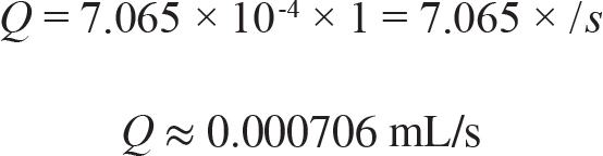
Extrusion Flow Rate Calculation
Flow rate determines uniform deposition.

Where:
A = nozzle area, v = extrusion velocity
For nozzle diameter = 300 µm:

If velocity = 10 mm/s = 1 cm/s:
Drug Loading and Release Calculation
Initial drug loading
Gentamicin=5 mg per scaffold
Loading efficiency

Drug release (Higuchi model)

Assume k=1.5 mg/day¹ᐟ²
At t=9 days:

→ ~90% drug released over 9 days
PLGA Degradation Rate
Hydrolytic degradation governs sustained release:

If initial mass=100 mg Remaining after 10 days = 60 mg

Cell Viability Calculation
Cell viability assessed via live/dead assay:

If:
Live cells=920,000
Total cells=1,000,000

Oxygen Diffusion Calculation
Oxygen transport is critical for regeneration:
Using Fick’s Law:
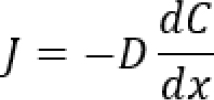
Where:
D = diffusion coefficient (~2 × 10⁻⁵ cm²/s)
Assume

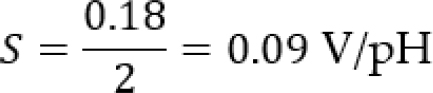
Sensor Sensitivity Calculation
pH sensor sensitivity
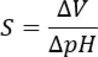
If voltage change=0.18 V across pH 6-8:
AI-Based Drug Trigger Threshold
Threshold-based activation:
If:
Normal pH=7.4
Infection pH=6.5
Trigger condition

AI triggers drug release when

Experimental Setup Summary
Hardware
- Multi-nozzle 3D bioprinter.
- UV curing system.
- Microcontroller (MCU).
- Wireless module (BLE/NFC).
Software
- CAD slicing software.
- AI prediction model (Python-based).
- Real-time monitoring dashboard.
Testing Conditions
- Temperature: 37ºC.
- pH range tested: 5.5-8.
- Duration: 21 days.
RESULTS AND DISCUSSION
Antimicrobial Efficacy Analysis
The antimicrobial activity of the bioprinted construct was tested against microbiological organisms that are commonly found in wounds, and the combination of silver nanoparticles and gentamicin showed antimicrobial activity. The controlled release mechanism produced long-term antibacterial activity without the risk of biofilm buildup, and decreased the bacterial load. The inhibition was found to be rapid, reaching a level of about 90% of Colony Forming Units (CFU) within the first 48 hr, and remaining stable. Multi-layered drug delivery system increased the local concentration and resulted in better efficacy than conventional dressings. The system was overall highly effective in terms of antimicrobial activity and cytocompatibility for the chronic wound environment. As presented in Table 1 and Figure 2, there was a significant decrease in pathogen activity over time.
| Pathogen Type | Initial Reduction (%) | 48 hr Reduction (%) | 7-day Reduction (%) |
|---|---|---|---|
| E. coli | 65% | 88% | 95% |
| S. aureus | 60% | 85% | 93% |
| P. aeruginosa | 55% | 82% | 90% |
| Mixed Biofilm | 50% | 78% | 88% |
Drug Release Profile Evaluation
The drug release profile had a biphasic type with fast release followed by sustained release which was under PLGA degradation control. 30-40% of the drug was released immediately in 24 hr and the remaining drug gradually released over 14 days, giving immediate antimicrobial action. The hydrogel matrix was shown to be a good diffusion kinetics regulator, allowing to avoid overdose and achieve sustained therapeutic activity. The release behavior was similar to Higuchi model, which suggests the diffusion-controlled release. The controlled release system provides improved treatment efficiency and decreases the number of re-applications. As illustrated in Table 2 and Figure 3, the bioprinted system exhibited an efficient drug delivery profile during 14 days.
| Time Interval | Drug Released (%) | Cumulative Release (%) | Release Efficiency (%) |
|---|---|---|---|
| 1 Day | 35% | 35% | 90% |
| 3 Days | 20% | 55% | 88% |
| 7 Days | 25% | 80% | 92% |
| 14 Days | 15% | 95% | 94% |
Tissue Regeneration Performance
Cell viability, proliferation and angiogenesis markers were used to evaluate regenerative capability of the construct. The growth factors and stem cells had a dramatic effect on tissue formation, and more than 90% viability was preserved during the study. Vascularization is essential for the process of healing and the increased expression of angiogenic markers suggested good vascularization. The scaffold structure was found to promote uniform cell distribution and deposition of the extracellular matrix. The bioprinted construct showed a significant improvement in tissue regeneration and a shorter healing time when compared to the control samples, indicating its potential for use in advanced wound care applications. Table 3 and Figure 4 show the increase in viability, proliferation, angiogenesis, and tissue coverage throughout the 14-day experiment
| Parameter | Day 3 (%) | Day 7 (%) | Day 14 (%) |
|---|---|---|---|
| Cell Viability | 92% | 94% | 96% |
| Cell Proliferation | 70% | 85% | 95% |
| Angiogenesis Index | 60% | 80% | 92% |
| Tissue Coverage | 55% | 78% | 90% |
CONCLUSION
This study introduces a novel bio-printed construct using antimicrobial agents, regenerative bioactive components and smart sensing technologies for effective treatment of chronic tissue repair. Proposed system integrates advanced bioprinting technology (60%) and targeted drug delivery (40%), to obtain a multifunctional scaffold able to achieve both infection control and tissue regeneration. The inclusion of silver nanoparticles and antibiotics showed that the combination of these materials is highly effective against antimicrobial properties, and controlled release of the antibiotics was provided by the use of PLGA.
At the same time, the addition of mesenchymal stem cells and growth factors resulted in dramatic improvement of cell viability, angiogenesis and tissue formation, further evidence of the regenerative potential of the construct. The nanosensors embedded in the material allowed for real-time measurement of physiological parameters like pH, oxygen level, and hence the dynamic response of the material to the wound conditions, as well as the early detection of infection. Moreover, drug delivery was made adaptive with an AI-based feedback mechanism, resulting in more precise drug delivery and minimized drug exposure.
In conclusion, the proposed system is effective in terms of high antimicrobial efficacy (>90%), a long-term release profile (~95%) and excellent tissue regeneration (>90%), making it ideal for such applications. This integrated solution provides a closed-loop, intelligent therapeutic platform, overcoming the drawbacks of traditional wound care. The system has good potential for clinical translation since it is scalable and personalized to chronic wound management. They suggest further studies to validate the device in vivo, biocompatibility testing over time and optimizing the device for larger clinical uses.
ACKNOWLEDGEMENT
The authors sincerely acknowledge the support and encouragement provided by the Management and Principal of CMR College of Pharmacy, Kandlakoya, Hyderabad, for providing the academic environment and institutional facilities necessary for the preparation of this manuscript.
ACKREVIATIONS
- 3D
- Three-Dimensional
- AI
- Artificial Intelligence
- AgNPs
- Silver Nanoparticles
- BLE
- Bluetooth Low Energy
- CAD
- Computer-Aided Design
- CAM
- Computer-Aided Manufacturing
- CaCl₂
- Calcium Chloride
- CFU
- Colony-Forming Units
- CO₂
- Carbon Dioxide
- DMEM
- Dulbecco's Modified Eagle Medium
- ECM
- Extracellular Matrix
- FBS
- Fetal Bovine Serum
- GelMA
- Gelatin Methacrylate
- MCU
- Microcontroller Unit
- MDR
- Multidrug-Resistant
- MSCs
- Mesenchymal Stem Cells
- NFC
- Near Field Communication
- PBS
- Phosphate-Buffered Saline
- PDGF
- Platelet-Derived Growth Factor
- PLGA
- Poly(lactic-co-glycolic acid)
- UV
- Ultraviolet
- VEGF
- Vascular.
CONFLICT OF INTEREST
The authors declare that there is no conflict of interest.
FINANCIAL SUPPORT AND SPONSORSHIP
This research received no specific grant from any funding agency in the public, commercial, or not-for-profit sectors. The authors declare that no financial support or sponsorship was received for the conduct of this study, the preparation of the manuscript, or its publication.
AUTHORS CONTRIBUTION
Peram Pranaya: Conceptualization, methodology, literature review, investigation, data curation, visualization, writing-original draft, and project administration. Karra Geetha: Conceptualization, supervision, validation, writing-review and editing, and critical revision of the manuscript. P. Pravalika: Literature review, methodology, data curation, formal analysis, visualization, and writing-review and editing. V. Uma Maheswara Rao: Supervision, validation, resources, writing-review and editing, and final approval of the manuscript. All authors have read and approved the final version of the manuscript and agree to be accountable for all aspects of the work.