
Article · Volume 17, Issue 1 · pp. 440–447
Therapeutic Efficacy of an Ethyl Pyruvate-Loaded Emulgel in Accelerating Wound Healing
Fareeda Bhanu Shaik1, Sudheer Akkiraju1, Zainab Tabassum Mohammad1, Golla Harshitha2, Sake Haritha2, Toluchuru Gamya Sree2, Kiran Sai Maccha2, Mahesh Vaggu2, Nagashubha Bobbarjang2, Somasekhar Reddy Kanala1
- 1Department of Pharmacology, Raghavendra Institute of Pharmaceutical Education and Research (RIPER)-Autonomous, K R Palli Cross, Ananthapuramu, Andhra Pradesh, INDIA.
- 2Department of Pharmaceutics, Raghavendra Institute of Pharmaceutical Education and Research (RIPER)-Autonomous, K R Palli Cross, Ananthapuramu, Andhra Pradesh, INDIA.
Abstract
Background: Ethyl Pyruvate (EP), is an ester of pyruvate with amazing anti-inflammatory, antioxidant, and antimicrobial properties. But EP’s poor aqueous solubility hinders its topical transport. To formulate an EP-loaded emulgel and evaluate its wound-healing efficacy in a rat excisional wound model.
Materials and Methods: An oil-in-water emulsion containing EP was prepared using cotton seed oil, Tween 80/propylene glycol, Span 20, propylene glycol and carbomer as the gelling agent. The emulgel became characterised for droplet size, polydispersity index, pH, viscosity, spread ability, and EP content. Full-thickness 2 cm dorsal excisional wounds were created in male Wistar rats. Animals (n=6 per group) grouped as control, blank gel, silver sulfadiazine and EP emulgel (2% & 4% w/v doses) and them treated daily for 14 days. Wound healing markers such as percentage wound contraction, oxidative stress markers in wound tissue include catalase, reduced glutathione, superoxide dismutase and malondialdehyde and the inflammatory marker myeloperoxidase, were measured. In addition, histopathology of wound tissue was performed.
Results: The optimized EP emulgel exhibited an average globule size of 280±0.21nm with Polydispersity Index (PDI) of 0.3, pH 6.54±0.17, viscosity 9521±0.07 cP and uniform EP content (>98%). EP treated wounds showed dose dependent improvement in wound contraction with p < 0.01 and 0.001 at day 7 (66% vs. 34%) and day 14th (95% vs. 70%) compared to controls. EP emulgel markedly reduced tissue Malondialdehyde (MDA), Myeloperoxidase (MPO) and the same time as restoring Superoxide Dismutase (SOD), Catalase (CAT) and reduced Glutathione (GSH) activity toward normal. Histology of skin revealed stronger re-epithelialization, denser collagen networks, and reduced inflammatory infiltrate inside the EP group.
Conclusion: EP-loaded emulgel efficiently promotes the wound-healing process, as evident by significant wound contraction, which may be due to its antioxidant and anti-inflammatory effects at wound site.
Keywords: Anti-inflammatory activity; Antioxidant activity; Ethyl Pyruvate Emulgel; Topical Formulation; Wound Healing
FULL TEXT
INTRODUCTION
The process of wound healing involves a series of events starting with tissue inflammation, tissue formation, and tissue remodelling (Fernández-Guarino et al., 2023). These processes work together to restore the integrity and function of damaged skin. However, factors such as infection, excessive inflammation, oxidative stress, and poor blood circulation can delay the healing process and lead to chronic wounds (Liu et al., 2022). Hence, need to develop effective topical formulations which accelerate wound repair and reduce complications has become an important area of pharmaceutical research (Tiwari & Pathak, 2023). In recent years, natural and bioactive compounds having to suppress tissue inflammation and oxidative tissue injury gained best choice of wound management strategies (Huang et al., 2022). Ethyl pyruvate has been widely reported as an anti-inflammatory, antioxidant, and cytoprotective effects (Yadav & Nair, 2025). These properties make it suitable as a promising agent for wound-healing activity. By minimizing oxidative damage and controlling inflammatory responses, ethyl pyruvate may enhance the overall wound healing process (Babalhakam et al., 2025). Topical drug delivery systems play a vital role in wound care because they allow direct contact of the therapeutic agent to the affected area (Zhao et al., 2023). Emulgel has become a cutting-edge and successful topical product. Emulgel combines the properties of both emulsions and gels, providing improved stability, enhanced drug release, and better patient compliance (Mohapatra et al., 2025). It is particularly useful for incorporating hydrophobic drugs into a gel base, thereby improving their penetration and therapeutic effectiveness (Slavkova et al., 2023). The incorporation of ethyl pyruvate into an emulgel formulation may offer several advantages in wound treatment. The gel base provides ease of application and prolonged contact with the skin, while the emulsion deliver its active principles efficiently at the wound site(Hashempur et al., 2025). This combination may enhance the therapeutic activity of ethyl pyruvate and support faster wound closure (Lu et al., 2021). Therefore, the present study focuses on the development and evaluation of ethyl pyruvate loaded emulgel and its potential wound healing effect (Ding et al., 2024). The aim is to explore whether this formulation can serve as an effective topical treatment for promoting wound healing and improving the overall recovery process (Ribeiro et al., 2024).
MATERIALS AND METHODS
Chemicals used in the study
Ethyl pyruvate (CAS 617-35-6) was procured from Otto Chemie Pvt. Ltd., Carbopol was obtained from Loba Chemie, while propylene glycol was purchased from SD Fine-Chem Limited. Triethanolamine was supplied by Qualikems. Tween 80, Span 20, methylparaben, propylparaben, and Isopropyl Myristate (IPM) were obtained from Merck. Marketed silver sulfadiazine was procured from local medical shop.
Pre-formulation studies
A solution containing 10-100 µg/mL of ethyl pyruvate was made using ethanol or phosphate buffer (pH 7.4). Absorbance values were recorded at a wavelength of 288 nm. A calibration curve plotting absorbance versus concentration was generated.
Emulgel Formulation Process
The emulgel was prepared by three steps that includes
Step 1: Emulsion Formation
The aqueous phase was prepared by mixing Tween 80 (15 mL) with 50 mL of water with constant stirring (800 rpm; 70ºC) for 30 min to ensure proper blending of the ingredients. After heating, the solution was allowed to cool gradually with continued stirring for another 20 min to maintain uniformity. Separately, the oil phase was prepared by combining Span 20, propylene glycol (10 mL), and cottonseed oil (5 mL) under heating (70ºC) with continuous stirring (800 rpm) for 30 min to obtain a clear and homogeneous product. After that, ethyl pyruvate (2% w/v) was mix with oil phase with continuous stirring. Now oil phase was slowly dropped into the aqueous solution with constant stirring (1 hr) at 800 rpm up to formation of a stable emulsion with uniform consistency. Similarly, ethyl pyruvate (4%w/v) nano emulsion was developed.
Step 2: Gel Base Preparation
Aqueous gel was prepared with carbopol 940 i.e. (0.85%w/v) under magnetic stirrer. The pH consistency of gel was adjusted with triethanolamine.
Step 3: Formation emulgel
The final prepared emulsion was made to gel by adding the gel base under continuous stirring.
Step 4: Characterization
The obtained formulation was characterized for the pH, viscosity, drug content and globule size (Karami et al., 2023).
Animal Grouping and Housing Conditions
Before the study, protocol was approved and handled as per CCSEA guidelines with following approval number: IAEC/VII/03/RIPER/2025.
Experimental design (n=6 Male Wistar rats/group)
| Group | Treatment |
|---|---|
| I | control |
| II | Plain emulgel |
| III | Standard; silver sulfadiazine (1% w/v) |
| IV | Ethyl pyruvate loaded emulgel (2% w/v) |
| V | Ethyl pyruvate loaded emulgel (4% w/v) |
Wound Healing Study (Excisional Model)
A circle skin wound with 2 cm diameter was created on dorsal sides of each rat under slight anaesthesia by injecting ketamine (50 mg/kg) and xylazine (10 mg/kg) intraperitoneally by complete removal of localised skin on the shaved dorsal region. The test formulation was applied on surface of wound daily up to 14 days. In between determine the quantify the wound closure by measuring diameter meter of wound at regular intervals at the end of protocols (Shailajan et al., 2011).

Biochemical Parameters
Preparation of Skin Tissue for Biochemical Assessment
After the 14-day treatment, wound skin tissue was carefully excised under ketamine plus xylazine anaesthesia, washed properly with saline (cold) and prepare tissue homogenate (10% w/) under cold condition and then utilised to estimate inflammation and oxidative stress markers (Halim et al., 2024).
Catalase (CAT) content
Catalase was determined in wound tissue homogenate as per earlier established Claiborne (1985) method. In a falcon tubes add phosphate buffer (2.5 mL), H₂O₂ (0.2M, 0.4 mL), and homogenate (0.1 mL) sequentially and mix uniformly. The absorbance was noted at 240 nm at 30-sec intervals over a 3-min period (Shalavadi et al., 2013).
Calculation
Quantify the catalase activity as per the following

Reduced Glutathione (GSH) Content
Wound tissue GSH content was determined as per the protocols of Ellman (1959) method. Add trichloroacetic acid (10%v/v) to the tissue homogenate and subjected for centrifugation to get clear supernatant. To this, add of DTNB reagent (0.5 mL) and phosphate buffer (3 mL, 0.2 M, pH 8.0). The solution absorption was recorded at 412 nm using a spectrophotometer. GSH concentration was calculated using its molar extinction coefficient (1.36 × 10⁴ M⁻¹ cm⁻¹) (Raza et al., 1991).
Calculations

Superoxide Dismutase (SOD) content
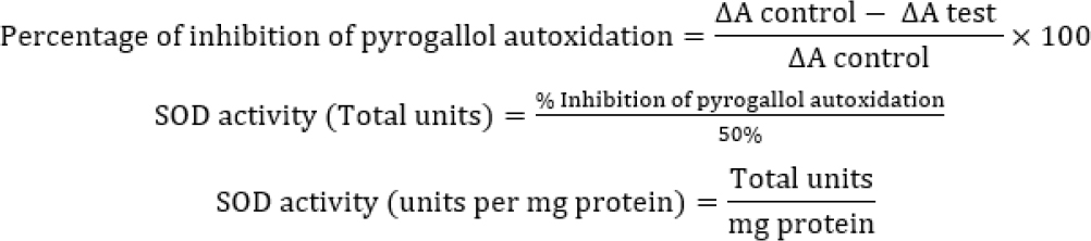
Determined the SOD activity as per Marklund and Marklund (1974) procedure. As per procedure, pyrogallol (10 mM) was added to supernatant (100 µL) at alkaline pH (8.4). The absorbance of this solution was recorded at 420 nm every 30 sec for a total duration of 3 min. The enzyme activity was calculated as per the equation provided (Marklund & Marklund, 1974).
Malondialdehyde (MDA) Levels
The amount of MDA was determine by the Buege and Aust (1978). As per the protocol, in the falcon tubes add wound tissue homogenate, TCA (15% w/v), TBA (0.375%w/v), and HCl (5N) sequentially, keep at 95ºC for 20 min and then centrifuged after attaining room temperature. The OD of supernatant was recorded at 532 nm. The concentration of lipid peroxides was calculated as per the give formula (Buege & Aust, 1978).

Myeloperoxidase levels
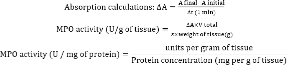
Granulation tissue was homogenized using HTAB solution made with phosphate buffer and was centrifuged. The collected supernatant was added with o-dianisidine solution and H₂O₂. The absorbance of resulting solution was noted using a spectrophotometer at 460 nm (Hanning et al., 2023).
Calculation
Histopathological Examination
On Day 14, the isolated wound skin was dehydration with alcohol and implanted in a paraffin block and was cut into small sections (five microns) and then stained with H&E.
Data analysis
The data was is expressed as Mean±SEM (n=6). Statistical evaluation of the data was carried out using GraphPad Prism version 10.2.1. One-Way Analysis of Variance (ANOVA), followed by Dunnett’s multiple comparison test are used to confirm the statistical difference among the groups. A p-value of less than 0.05 was considered statistically significant.
RESULTS
UV-Spectrometric Method
The analytical method for ethyl pyruvate was performed using UV-visible spectrophotometric method at 288 nm. It obeyed Beers-Lambert law’s range of 10-100 μg/mL. The obtained R2 value of 0.9951 indicates that the calibration curve was linear and confirms the effectiveness of the drug content evaluation (Figure 1).
Characterization of Emulgel
Globule size
Globule size was estimated using the Nano sizer (Horiba SZ-100). The globule size of the ethyl pyruvate loaded emulgel was found to be 280±0.21 nm with PDI of 0.390 (Figure 2).
pH and Viscosity
The pH of the Emulgel formulation was found to be 6.54±0.17. The viscosity of the Ethyl pyruvate loaded emulgel was found to be 9521±0.07cP (Table 1).
| Parameter | Ethyl pyruvate loaded Emulgel |
|---|---|
| pH | 6.54±0.17 |
| Viscosity | 9521±0.07 cP |
Values are represented as Mean±SEM; cP means centipoise.
Determination of drug content
The amount of ethyl pyruvate in the prepared emulgel ranged from 98.54±0.21% to 99.54±0.36%. The results established the consistent distribution of ethyl pyruvate within the emulgel matrix with negligible loss during formulation.
Wound healing activity
Wound contraction was non-significant at day 01 in all the groups, indicating uniform wound induction. However, from Day 3 onwards, EP emulgel groups exhibited a significant increase in wound closer than control. Notably, on 7th Day, 4%w/v EP emulgel was demonstrated greater wound contraction than 2%w/v dose (p<0.05), confirming a dose-dependent improvement in wound healing process. On 14th day, EP (2%w/v & 4%w/v) treated groups showed with highest wound contraction (>90%) compared to control (70%) (Table 2 and Figure 3).
| Groups | Day | |||
|---|---|---|---|---|
| 1 | 3 | 7 | 14 | |
| I | 5.10±0.8 | 21.70±0.5 | 34.45±0.57 | 70.00±0.18 |
| II | 5.60±0.7 ns | 26.20±0.80 * | 40.80±0.82 ** | 81.20±0.30 ** |
| III | 5.90±0.6 ns | 55.10±0.48 *** | 72.20±0.63 *** | 99.10±0.18 *** |
| IV | 5.70±0.9 ns | 38.80±0.86 *** | 58.20±0.90 *** | 92.70±0.84 *** |
| V | 6.00±0.7 ns | 45.90±0.42 ### | 66.80±0.70 ###† | 95.20±0.56 ###† |
Significant p values were considered as fallows less than 0.05 values with *; 0.01 values with **; 0.001 *** and † p<0.05 vs Group IV (dose-dependent significance).
Effect of Ethyl pyruvate emulgel on wound tissue markers
Treatment with ethyl pyruvate produced a clear dose-dependent improvement in oxidative stress parameters when compared with control and plain gel groups. The ROS-neutralising factors like superoxide dismutase, reduced glutathione, and catalase were significantly decreased in the control group, while treatment with ethyl pyruvate, particularly at 4% w/v, markedly elevated in the wound tissue (p-value <0.001), approaching or matching with silver sulfadiazine group, indicating restoration of endogenous antioxidant defence. In contrast, markers of oxidative damage and inflammation, namely Malondialdehyde (MDA) and Myeloperoxidase (MPO), were significantly elevated in the control group and remained unchanged in topical placebo gel applied group confirming the absence of therapeutic effect of the base. However, Topical application of EP-loaded emulgel markedly reduced lipid peroxidation and myeloperoxidase (p<0.01 to p<0.001), with the 4% formulation showing greater reduction, in some cases comparable to or better than the standard. Overall, these findings prove that ethyl pyruvate effectively attenuates wound tissue oxidative damage and inflammation by enhancing antioxidant defence system and neutrophil infiltration, with superior efficacy observed at higher concentration (Figure 4).
Histopathological studies
Histopathological evaluation revealed marked transformations among the experimental groups, indicating progressive tissue repair with treatment. The control group exhibited disrupted epidermal architecture with irregular epithelial lining and loosely arranged collagen fibers, indicating impaired healing. The plain gel group showed similar histological features with mild improvement, characterized by incomplete re-epithelialization, confirming its minimal therapeutic effect. In contrast, the standard-treated group demonstrated well-organized epidermis with complete re-epithelialization and restoration of normal skin appendages, indicating effective wound healing. The low-dose ethyl pyruvate (2%) group showed moderate healing with partial re-epithelialization whereas the high-dose ethyl pyruvate (4%) group exhibited near-complete restoration of skin architecture, with a well-formed epidermal layer, dense and organized collagen bundles and improved dermal structure, closely resembling the standard group (Figure 5).
DISCUSSION
In this study, the ethyl pyruvate-loaded emulgel exhibits significant and dose-dependent enhancement of wound healing, which can be attributed to its combined physicochemical suitability and biological activity. The formulation showed optimal characteristics, including a near-neutral pH (6.54±0.17) compatible with skin physiology, appropriate viscosity (9521±0.07 cP) ensuring good retention and spreadability, nanosized globules (~280 nm) and PDI of 0.390 facilitating enhanced skin penetration, and high drug content uniformity (~98-99%), collectively indicating an efficient topical delivery system (Das et al., 2025). These add up qualities expected to improve therapeutic outcomes during animal testing. The percentage of wound closure was higher, particularly with the 4% w/v formulation, which achieved ~99% contraction on day 14th but only attain ~70% wound closure in the control, confirming a strong dose-dependent response. The reason for better wound healing effect may be associated with increased fibroblast proliferation and myofibroblast-mediated wound closure, which are critical for tissue remodelling (Morris et al., 2025). Furthermore, biochemical analysis showed that ethyl pyruvate significantly restored antioxidant defence systems, as evidenced by increased levels of SOD, CAT, and GSH, alongside a marked reduction in lipid peroxidation (MDA) and inflammatory marker MPO. This indicates that the formulation effectively mitigates oxidative stress and inflammation, both of which are known to delay wound healing if uncontrolled (Wang et al., 2023). The histopathological findings further corroborated these results, showing improved re-epithelialization, organized collagen deposition, and reduced inflammatory infiltration in treated groups, with the 4% formulation closely resembling the standard drug (silver sulfadiazine), thereby confirming enhanced tissue regeneration. Mechanistically, ethyl pyruvate has proven compound for its anti-inflammatory and antioxidant effects, which help regulate the inflammatory phase and prevent prolonged tissue damage, thereby promoting faster progression to the proliferative and remodeling phases of healing (Yang et al., 2016). Additionally, the emulgel system itself enhances drug bioavailability and penetration across the stratum corneum, supporting sustained drug release and improved therapeutic efficacy (Donthi et al., 2023). Overall, the findings suggest that the ethyl pyruvate emulgel not only accelerates wound contraction but also improves the quality of curing through modulation of oxidative stress, inflammation.
CONCLUSION
The emulgel loaded with ethyl pyruvate has shown beneficial effects on wound healing due to antagonise the wound tissue damage by the ROS, inflammation and encourage epithelial tissue regeneration upon topical application. For approved clinical indication need more research could be necessary.
ACKNOWLEDGEMENT
The authors are thankful to principal, management and the DST-FIST facility of Raghavendra Institute of Pharmaceutical Education and Research (RIPER) for providing the necessary facilities for carrying out this project.
ABBREVIATIONS
- CCSEA
- Committee for the Purpose of Control And Supervision of Experiments on Animals
- H₂O2
- Hydrogen Peroxide
- DTNB
- 5, 5′-dithiobis (2-nitrobenzoic acid
- EP
- Ethyl pyruvate
- SEM
- Standard Error of the Mean
- MPO
- Myeloperoxidase
- SOD
- Superoxide Dismutase
- CAT
- Catalase
- GSH
- Reduced Glutathione
- MDA
- Malondialdehyde
- ANOVA
- Analysis of variance
- H&E
- Haematoxylin and Eosin staining
- HTAB
- Hexadecyl Trimethyl Ammonium Bromide
- TBA
- Thiobarbituric Acid
- OD
- Optical Density
- TCA
- Trichloroacetic acid
- ROS
- Reactive oxygen species
- PDI
- Polydispersity Index
- IPM
- Isopropyl myristate.
CONFLICT OF INTEREST
The authors declare that there is no conflict of interest.
ETHICAL STATEMENT
The research protocol was approved by Institutional ethical committee of Raghavendra Institute of Pharmaceutical Education and Research (RIPER)-Autonomous, Ananthapuramu, Andhra Pradesh with following approval number: IAEC/VII/03/RIPER/2025.