
Article · Volume 19, Issue 1 · pp. 176–184
Chemically Synthesized Urolithin-C Regulates Glucose Uptake in HepG2 Cells
Vani Manjappa Karadi1,2, Manjula Mavathuru Venkatappa3, Shivakumar Venkataramaiah3, Sanjana Bai Shivaram Singh Mahendaranath Singh1, Sushma Mohan1, Ramesh Komalapura Lakshmaiah4, Devaraja Sannaningaiah1,3
- 1Department of Studies and Research in Biochemistry, Tumkur University, Tumakuru, Karnataka, INDIA.
- 2Department of Biochemistry, Maharani Lakshmi Ammanni College for Women, Bengaluru, Karnataka, INDIA.
- 3Department of Studies and Research in Food Science and Nutrition, Tumkur University, Tumakuru, Karnataka, INDIA.
- 4Home Science, Maharani’s Science College for Women, Mysuru, Karnataka, INDIA.
Abstract
Background: In recent years, the prevalence of diabetes has increased dramatically worldwide. Although several drugs are available for its management, their severe cytotoxic effects have created a need for safer and more effective alternatives. Urolithins (A, B, C, D, M5, M6, and M7) have gained attention due to their pharmacological potential; however, Urolithin-C remains the least studied. In our previous study, we reported the anti-inflammatory mechanism of chemically synthesized Urolithin-C.
Objectives: In this current study, the effect of chemically synthesised Urolithin-C on glucose uptake in HepG2 cells was examined.
Materials and Methods: The antidiabetic activity of synthetically synthesised Urolithin-Con was assessed by measuring its inhibitory effects on α-amylase, α-glucosidase, and dipeptidyl Peptidase-IV (DPP-IV), along with glucose uptake studies in HepG2 cells.
Results: The inhibitory effect of Urolithin-C on α-amylase activity was shown by 57.14% (IC₅₀=352.69 μg/mL) and α-glucosidase activity by 64.40% (IC₅₀=296.83 μg/mL) compared with the positive control acarbose. Urolithin-C in a dose-dependent manner inhibited DPP-IV enzyme activity by approximately 68% compared with sitagliptin (72%). Molecular docking analysis of α-amylase and α-glucosidase showed binding energies of -7.7 kcal/mol and −8.2 kcal/mol, respectively with the active sites. Later, Urolithin-C enhanced glucose uptake by 58.24% in HepG2 cells after 4 hr of exposure, measured through 2-NBDG fluorescence intensity. Metformin (100 µM/mL) was used as the standard control and stimulated 64.53% by flow cytometry.
Conclusion: Urolithin-C demonstrates promising antidiabetic potential by enhancing the activity of the antioxidant enzyme G6PD, as indicated by a cellular G6PD assay.
Keywords: α-amylase; α-glucosidase; Anti-diabetic Activity; Dipeptidyl Peptidase-IV; Urolithin-C
FULL TEXT
INTRODUCTION
Diabetes Mellitus (DM) is a chronic metabolic disorder that occurs when the pancreas fails to produce sufficient insulin or when the body rejects the utilisation of produced insulin, resulting in persistent hyperglycemia. The β-cells of the pancreas secrete insulin, a peptide hormone that regulates blood glucose levels (Tafesse et al., 2017). In the absence of adequate or effective insulin action, the level of blood glucose increases, leading to long term damage to the vital organs such as the heart, kidneys, eyes, nerves, and blood vessels. Hence, diabetes mellitus is a multifactorial metabolic disorder characterised by disturbances in carbohydrate, fat, and protein metabolism due to impaired insulin secretion/insulin resistance (both). It is also associated with elevated levels of oxidative stress, inflammation, and pancreatic β-cell dysfunction (Guerra et al., 2021). Diabetes mellitus represents a growing global health burden, affecting over 830 million people worldwide (approximately 14% of adults), with 90-95% being Type 2 diabetes. Terrifyingly, nearly 40-45% of cases remain undiagnosed, and projections estimate that the number of affected individuals may exceed 850 million by 2050, particularly in low and middle-income countries (Yameny, 2024). Under physiological conditions, glucose homeostasis is maintained through tightly regulated insulin signalling pathways. The binding of insulin to its tyrosine kinase receptor triggers autophosphorylation and activation of insulin receptor substrates (IRS-1 and IRS-2), which subsequently stimulate the PI3K/Akt pathway. This cascade promotes glucose uptake through translocation of GLUT-4 transporters to the cell membrane and suppresses gluconeogenic gene expression (Derosa and Maffioli, 2012). Disruption of these pathways contributes significantly to the development of diabetes.
Currently, the management strategies include increased physical activity, weight management, balanced nutrition, and pharmacological interventions. For Type 1 diabetes, insulin therapy is essential, while Type 2 diabetes management focuses on improving insulin sensitivity and controlling postprandial hyperglycemia. One therapeutic strategy involves inhibiting the breakdown of dietary carbohydrates (starch, sugars, and fibres) into absorbable monosaccharides (Parmar et al., 2012; Gupta et al., 2009). Enzymes such as α-amylase, α-glucosidase and DPP-IV play a crucial role in carbohydrate digestion into glucose, which is then absorbed into circulation (Alberti and Zimmet, 1998). Therefore, inhibition of these enzymes delays glucose absorption and helps to reduce postprandial blood glucose levels. Acarbose, voglibose and miglitol inhibits the activity of α-amylase and α-glucosidase, while sitagliptin, vildagliptin, sitagliptin and linagliptin inhibits the activity of DPP-IV. The said inhibitors despite their glucose-regulating potential, cause gastrointestinal discomfort such as flatulence, diarrhoea, and abdominal pain due to colonic fermentation of undigested carbohydrates (Srinivasan et al., 2017; Kim and Egan, 2008). Consequently, there is a need for safer and more effective therapeutic alternatives (Prigeon et al., 2003; Ball et al., 2002).
Fruits such as pomegranate, berries, walnuts and almonds are the storehouse of Ellagitannins (ET), a group of polyphenolic compounds (Dall’Asta et al., 2015). ETs in the small intestine undergo degradation by gut microbiota into ellagic acid later converted into various forms of pharmacologically active urolithin derivatives (Leng et al., 2025). Among the said urolithin A and B have been extensively researched as they have been found to exhibit antioxidant, anti-inflammatory, antidiabetic and metabolic regulatory effects (García-Villalba et al., 2022). Urolithin A activates the AMPK pathway, improves insulin sensitivity, enhances glucose uptake in muscle and liver cells and regulates mitochondrial quality (Bai et al., 2017). While Urolithin B improves glucose metabolism, reduces lipid accumulation, exhibits weaker anti-inflammatory and antidiabetic activity than Urolithin A (Chen et al., 2024). Urolithin-C is one of the least studied metabolites of the Urolithin family. In our previous study, we reported the antioxidant and mechanism of anti-inflammatory action of chemically synthesised Urolithin-C (Manjappa et al., 2025). Therefore, in the present study, we made an attempt to investigate the antidiabetic potential of chemically synthesised Urolithin-C and the results are presented.
MATERIALS AND METHODS
Chemicals and reagents
α-amylase from Aspergillus oryzae, NaH2PO4, Na2HPO4, Starch, DNS Reagent, α- glucosidase from Saccharomyces cerevisiae, p-Nitrophenyl-β-Glucopyranoside (pNPG), Sodium Carbonate, DPP-IV Drug discovery kit, Acarbose, HEPG2- Human hepatocellular adenocarcinoma cell line (NCCS, Pune, India), MEM-alpha medium, Fetal Bovine Serum, D-PBS, Metformin hydrochloride, DMSO, MTT, Antibiotic-Antimycotic solution-100x, Trypsin EDTA, Human Glucose-6 -Phosphate 1-dehydrogenase activity assay kit, were acquired from the Sigma Aldrich Corporation in St. Louis, Missouri, USA. Each chemical is of analytical quality.
Anti-diabetic activity
Inhibition of α-amylase activity
The activity of α-amylase was carried out using previously reported techniques (Alqahtani et al., 2019). Various concentrations of Urolithin-C (12.5-400 µg/mL) were used in dry test tubes and 96-well plates, respectively. The samples were preincubated with α-amylase enzyme solution (0.5%) prepared in 0.1 N phosphate buffer (pH 6.8), using a total volume of 500 µL in test tubes and 50 µL in microplate wells. The mixture was incubated at room temperature for 30 min. Following preincubation, an equal volume of 1% (w/v) soluble starch solution prepared in the same phosphate buffer (pH 6.8) was added to initiate the enzymatic reaction. The reaction mixture was further incubated at room temperature for 10 min. The reaction was terminated by adding freshly prepared 3,5-Dinitrosalicylic Acid (DNS) reagent. Subsequently, the reaction mixtures were heated in a boiling water bath at 90-95ºC for 8-10 min to facilitate colour development. After cooling to room temperature, the absorbance was measured at 540 nm using a microplate reader for the 96-well format and a spectrophotometer for test tube assays. Acarbose was used as the positive control. The % inhibition (I%) was calculated by using the following formula:

Ac -absorbance of the Control, As-absorbance of the Sample.
Inhibition of α-glucosidase activity
The α-glucosidase inhibition activity was carried out through the test tube and 96-well plate method, followed by previously described methods (Alqahtani et al., 2019; Elya et al., 2012). In brief, different concentrations of Urolithin-C (12.5-400 µg/mL) in dry-cleaned test tubes and 12.5-400 µg/mL in 96-well plates were taken and pre-incubated samples with 500 µL in a test tube and 50 µL in a 96-well plate of alpha-glucosidase enzyme (0.5 U/mL prepared using 0.02 M phosphate buffer) were added and incubated for 20 min at 37ºC. After that, an equal volume of the substrate p-nitrophenyl-α-D-glucopyranoside (pNPG -3 mM) was added to both and the reaction mixture was incubated for 10 minutes at 37ºC. Later, 150 µL of 0.1M Na2CO3 in a 96-well plate and 2 mL of 0.1 M Na2CO3 in a test tube were added to stop the reaction. Spectrophotometric and microplate readings were taken at 405 nm. Acarbose was used as a control for the study. The Percentage of α-glucosidase Inhibition (I%) was calculated by using the following formula:
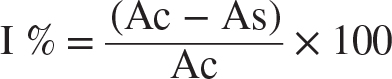
Ac-Absorbance of the Control, As-Absorbance of the Sample.
DPP-IV Inhibition Study
The DPP-IV enzyme inhibition activity was examined as followed by the previously described method (Amin et al., 2019). A clean dry 96-well plate was filled with a different concentration (12.5-400 µg/mL) of Urolithin-C and 30 µL of assay buffer, followed by the addition of inhibitor sitagliptin (p32/98 standard). Then 10 µL of DPP-IV enzyme was added to the well plate, except the blank (assay buffer alone) well and the plate was incubated for 10 min at 37ºC. After preincubation, 50 µL of substrate Gly-pro-pNA was added to the well plate and the reaction mixture was kept for 30 min at 37ºC. Then the absorbance was measured at 405 nm with the help of a 96-well plate reader. The DPP-IV enzyme inhibition of Urolithin-C was calculated using the following formula:

Molecular docking
The Urolithin-C compound and protein (PDB ID: 4W93 and 3TOP) were subjected to docking using Auto Dock Vina to position them within the active region of proteins. The chemical structure of the compound was obtained from the PubChem database with appropriate 2D orientation. Additionally, the molecule's energy was minimised using ChemBio3D. The ligand molecule, which had undergone energy minimisation, was subsequently employed as input for Auto Dock Vina to conduct the docking simulation. The crystallographic structure of the receptor molecules, amylase and glucosidase (Protein Data Bank (PDB) ID: 4W93 and 3TOP), was obtained from the PDB. The structures were modified by eliminating all heteroatom coordinates and water molecules and incorporating hydrogens, Kollman charges, and the absent C-terminal oxygen. The Auto Dock 4.2 program was designed to accept only receptor files that do not contain residues with non-integral charges. Subsequently, the target protein files were generated by excluding the related residue from the protein structure using the Auto Dock 4.2 auto-preparation of target protein files (MGL tools 1.5.6) (Kajaria et al., 2013).
Cell Line Maintenance
The cell line HepG2 (Human hepatocellular adenocarcinoma cell line) was purchased from NCCS, Pune, India. The purchased cells were maintained in Minimum Essential Medium (MEM) with 2 mM L-glutamine and Earle's balanced salt solution contained 1.5 g/L sodium bicarbonate, 0.1 mM non-essential amino acids and 1 mM sodium pyruvate supplemented with 10% Fetal Bovine Serum (FBS) along with the 1% antibiotic-antimycotic solution maintained in the modified atmosphere with 5% CO2 and 18-20% O2 in the CO2 incubator at 37ºC. After reaching 80% of density, cells were detached using 0.025% trypsin and 0.01% EDTA (in Dulbecco's Phosphate Buffered Saline) solution.
Cell viability and proliferation studies
MTT assay and cell morphological assessment
The cytotoxicity effect of Urolithin-C was measured on HepG2 cells through the MTT assay, followed by the previously published method (Odeyemi and Dewar, 2019a). In brief, 15,000 cells were plated in 200 μL of cell culture media was added to the 96-well plate and incubated at 37ºC for overnight. Later, the medium was replaced with DMEM having various concentrations of Urolithin-C (12.5-200 μg/mL). The cells were incubated for 24 hr at 37ºC with 5% CO2 atmosphere. After the incubation, the cells were treated with 100 μL of MTT reagent (0.5 mg/mL) and incubated for 3 hr at 37ºC. Then the formed formazan crystals were dissolved in 100 μL of DMSO and it was measured at 570 nm using a microplate reader. The viability of cells treated with DMEM alone was considered 100%. The percentage cell viability was calculated using the following formula:

The morphological changes in HepG2 cells treated with different concentrations (12.5 μg/mL to 200 μg/mL) of Urolithin-C for 24 hr at 37ºC. The treated cells were observed and recorded using an inverted biological microscope with a scale of 200 µM.
Glucose uptake study by Flow Cytometry: 2-NBDG staining
The Glucose uptake was analysed through flow cytometry by using 2-NBDG staining was carried out by using the method of Zou et al. (2005). Briefly, the cultured HepG2 cells were incubated in a CO2 incubator overnight at 37ºC with a cell density of 0.5 x 10⁶ cells/2 mL. Followed by the incubation, aspirate the spent medium and treat the cells with Urolithin-C (50 μg/mL) and metformin (100 μMol) as a positive control in 2 mL of glucose-free culture medium containing 100 μM 2-[N-(7-nitrobenz-2-oxa-1,3-diazol-4-yl)amino]-2-deoxy-D-glucose (2-NBDG) dye and keep the cells in dark conditions for 4 hr. Then removed the medium from the cells and washed the wells with Phosphate-Buffered Saline (PBS). Incubated cells at 37ºC for 3-4 min with 400 μL trypsin and cells were collected in 12 x 75 mm FACS tubes. Then, the tubes were centrifuged at 2700 RPM for 3 min at room temperature. The absorbance was measured at 540 nm.
Glucose-6 Phosphatase Dehydrogenase (G6PD) activity
The method of Li et al. (2013) was followed to determine the Glucose-6 Phosphatase Dehydrogenase (G6PD) activity. The cultured HepG2 cells in a 6-well plate at a density of 2 x 10⁵ cells/2 mL were incubated in a CO2 incubator at 37ºC for 24 hr. Followed by the above said Glucose uptake study protocol up to cell harvest, later the cells were collected and lysed and the lysate was used for further analysis. The cell lysate and standard metformin were pipetted into micro wells which contained Human Glucose-6-phosphate-1-dehydrogenase, G6PD presented in the samples, were bounded by the antibodies. A biotin-labelled antibody was added and followed by the addition of Streptavidin-HRP and incubated to form a complex. After microwells were washed using PBS and substrate solution 3,3′,5,5′-Tetramethylbenzidine (TMB) was added to the microwells and the developed colour was proportional to the amount of G6PD present in the treated cells. Then, the stop solution was added to stop the reaction. The absorbance is measured at 450 nm.
Statistical Analysis
The results were statistically evaluated and provided as mean±SD. Individual parameters were compared using one-way ANOVA in GraphPad Prism 5.0 (GraphPad Software, Inc., San Diego, CA, USA). The p-value (p<0.05) was considered as significant.
RESULTS
Urolithin-C inhibited the activity of α-amylase, α-glucosidase and DPPP-IV enzymes
The inhibition of α-amylase and α-glucosidase enzymes against the Urolithin-C was evaluated by test-tube and 96-well plate methods. Urolithin-C against the α-amylase and α-glucosidase in the test tube method showed a significant antidiabetic effect in a dose-dependent manner. In α-amylase, Urolithin-C inhibited 80.95% and the IC50 value was found to be 8.65 μg/mL. On the other hand, inhibition of α-glucosidase enzyme activity by Urolithin-C was found to be 78.1% with an IC50 value of 17.52 μg/mL. For both the enzyme activities, acarbose was used as a positive control drug and it was showed 86.76 % in α-amylase and 83.83% of inhibition in α-glucosidase (Figures 1A and 1B). Furthermore, the same enzymatic study was carried out by the 96-well plate method interestingly, it showed a satisfactory antidiabetic effect by inhibiting the α-amylase and α-glucosidase enzymes in a dose-dependent manner. About 57.14% of α-amylase inhibition was observed with an IC50 value of 352.69 μg/mL. The positive control acarbose showed about 98.62% inhibition with an IC50 value of 20.67 μg/mL (Figure S1A). On the other hand, Urolithin-C showed 64.40% of α-glucosidase inhibition with an IC50 value of 296.83 μg/mL, whereas about 97.81% α-glucosidase inhibition was examined in case of positive control acarbose with an IC50 value of 24.49 μg/mL (Figure S1B). In addition, Urolithin-C inhibited Dipeptidyl peptidase-IV (DPP-IV) activity in a dose dependent manner. Urolithin-C showed 68% DPP-IV inhibition with an IC50 value of 242.46 μg/mL Sitagliptin was used as a standard drug, showed 72% of inhibition (Figure 1C).
Urolithin-C interacts with histidine, aspartic acid, glutamic acid and arginine present in the active site of α-amylase, while glutamic acid and leucine are present in the α-glucosidase active site.
The interaction between the enzyme and inhibitor was investigated using molecular docking techniques. Urolithin-C showed binding energies of -7.7 kcal/mol and -8.2 kcal/mol when interacting with α-amylase and α-glucosidase, respectively (Figures S2 and S3). Urolithin-C engaged with the conserved amino acid residues HISA:101, ASPA:197, GLUA:233, ARGA:195 at the active site via hydrogen bonding of α-amylase enzyme, (GLUA:1284, LEUA:1291 of α-glucosidase interactions with the ligand Urolithin-C.
Urolithin-C exhibits a moderate cytotoxic effect on HepG2 cell lines
The cytotoxic effect of Urolithin-C on HepG2 cells was analysed by the MTT assay. The Urolithin-C was moderately toxic against the HepG2 cells, with the % cell viability of 72.85 % at the higher dose of 200 ug/mL after 24 hr of incubation. Metformin (100 µM) was used as a standard control against HepG2 cells, which showed 84.32 % viability. Based on the obtained cytotoxic results, 50 µg/mL was used for further mechanistic studies (Figure 2A). Furthermore, the morphological analyses of Urolithin-C-treated HepG2 cells were observed using an inverted microscope with a scale of 200 μM. While Urolithin-C at a greater concentration (200 µg/mL) had a smaller round body and a lower density in the treatment group, the morphology of HepG2 cells in the untreated group and metformin revealed a high density with adherent cell colonies (Figure B).
Urolithin-C significantly enhances glucose uptake in HepG2 cells
The role of Urolithin-C on glucose uptake in HepG2 cells was examined using 2-NBDG (2-(N-(7-Nitrobenz-2-oxa-1,3-diazol-4-yl) Amino)-2-Deoxyglucose), a fluorescent glucose analog. In case of untreated cells, 2-NBDG uptake was not noticed, suggesting the low basal glucose transport activity (Figure C). However, metformin-treated cells showed augmented glucose uptake with nearly about 65% of cells exhibiting positive 2-NBDG fluorescence. Similarly, in the case of Urolithin-C-treated HepG2 cells, there was an increase in glucose uptake, attainment nearly about 58%, slightly lower than the Metformin-treated group. In addition, flow cytometric analysis (Figure D) authenticated the observed results as well as untreated HepG2 cells showed a left-shifted peak, revealing low fluorescence intensity with least glucose uptake. On the hand, both Metformin and Urolithin-C treated cells displayed a rightward shift in fluorescence intensity, suggesting augmented accumulation of intracellular 2-NBDG. In comparison, metformin showed a prominent shift rather than Urolithin-C. Figure 3A indicates the dot plots of 2-NBDG fluorescence versus Side Scatter Signal (SSC). Relatively less 2-NBDG uptake was noticed in the case of untreated HepG2 cells as a lower fluorescence region was noticed. On the other hand, a moderate rise in fluorescence intensity was observed in Metformin, a standard drug treated cells. Interestingly, Urolithin-C treated cells showed rightward shift suggesting the enhanced uptake of 2NBDG. Figure B indicates the Forward Scatter (FSC) versus SSC plots endorsing uniform cell populations and proper gating across all experimental groups. Cells were distributed inside the gated region, consistently revealed the noticed difference in fluorescence was not due to a difference in cell size or granular nature rather the change in glucose uptake. Figure C represents the histogram overlays of quantitative glucose uptake distribution (2-NBDG fluorescence). Lower fluorescence intensity peak was noticed in the case of untreated group, while the positive control treated group displayed a shift towards higher fluorescence. Most importantly, the Urolithin-C-treated group showed a rightward shift in the histogram peak was observed suggesting the augmented buildup of 2-NBDG.
Urolithin-C enhances cellular antioxidant defense by upregulating G6PD activity
To ensure the cellular antioxidant potential of Urolithin-C, its effect on glucose-6-phosphate dehydrogenase (G6PD) activity was analysed (Figure 4). Basal G6PD level was found to be 8-9 ng/mL, however, metformin (Positive control) significantly raised the level of G6PD (15 ng/mL), suggesting the increased activity of the antioxidant enzyme. Urolithin-C modulated the G6PD activity in a dose dependent manner. At the concentration of 12.5 and 25 µg/mL, the level of G6PD was found to be increased (8-9 ng/mL), suggesting a significant improvement in comparison to the untreated group. Furthermore, at a concentration of 50 µg/mL of Urolithin-C, a marked increase in G6PD was observed (16-17 ng/mL), which was highly significant (p<0.001) compared with the metformin-treated group. However, at the concentration of (100 µg/mL) the level of G6PD was slightly lesser than 50 µg/mL. Most importantly, at the concentration of 200 µg/mL of Urolithin-C, the level of G6PD activity was significantly declined to ~10 ng/mL. These findings suggest that Urolithin-C through its antioxidant property, upregulates the activity of G6PD. The maximum effect was noticed at the concentration of 50 µg/mL. While declaimed activity at higher concentration (200 µg/mL) could be a possible biphasic effect dependent on the concentration.
DISCUSSION
The increasing frequency of diabetes mellitus globally opened the new avenues for the search for much safer yet effective therapeutic agents. Urolithins, a class of gut microbiota derived metabolites of ellagitannins, have been receiving significant evidence due to their immense pharmacological properties. Despite various types of Urolithins such as Uro-A, Uro-B, Uro-C, Uro-D, Uro-M5, Uro-M6 and Uro-M7 have been reported so far only Urolithin A and B have been extensively studied (Uzar et al., 2012). While Urolithin-C is a least explored member of this family which has emerged as a promising therapeutic agent. In our previous study we reported the antioxidant and mechanism of anti-inflammatory action of chemically synthesised Urolithin-C (Manjappa et al., 2025). In the current study, we examined the antidiabetic property of Urolithin-C, emphasising its capability to curb key carbohydrate-metabolising enzymes and to increase glucose uptake efficiency.
Diabetes mellitus characterised by elevated blood glucose levels, impaired insulin signalling, oxidative stress, and altered hepatic glucose metabolism (Unger and Parkin, 2010). Digestive enzymes such as α-amylase and α-glucosidase play critical roles in maintaining glucose homeostasis and are directly involved in postprandial hyperglycemia. Complex carbohydrates (polysaccharides such as starch) are first hydrolysed into oligosaccharides by α-amylase, and subsequently α-glucosidase converts carbohydrate intermediates into absorbable glucose units (Alam et al., 2022). Therefore, inhibition of these enzymes slows carbohydrate digestion and reduces intestinal glucose absorption. Interestingly, Urolithin-C exhibited fair inhibitory activity against both α-amylase and α-glucosidase similar to the standard inhibitor acarbose. These findings are consistent with previous reports describing the inhibitory potential of polyphenolic compounds such as ferulic acid, coumarin, 3-isopropylcatechol, and caffeine against carbohydrate-digesting enzymes (Odeyemi and Dewar, 2019b; Zhao et al., 2015).
In addition, the inhibitory effect of Urolithin-C on α-amylase and α-glucosidase was more authenticated by the molecular docking studies. The Urolithin-C showed a binding energy of -7.7 kcal/mol and -8.2 kcal/mol when it interacted with the α-amylase and α-glucosidase, respectively. Furthermore, Urolithin-C showed significant inhibition of Dipeptidyl Peptidase-IV (DPP-IV) enzyme that regulates the incretin signalling pathway. DPP-IV degrades incretin hormones including Glucagon-Like Peptide-1 (GLP-1) and Gastric Inhibitory Polypeptide (GIP), which enhance glucose-dependent insulin secretion and suppress glucagon release. Inhibition of DPP-IV is known to lower glucagon levels, delay gastric emptying, and promote insulin secretion (Alongi and Anese, 2018). Notably, Urolithin-C exhibited 68% DPP-IV inhibition compared to standard drug sitagliptin (P32/98) 72% inhibition was noticed; hence this indicated the substantial inhibitory effect on diabetes-associated enzymes.
Additionally, to assess cellular toxicity and metabolic competence of Urolithin-C was evaluated using an in vitro hepatic cell model (HepG2 cells). These cells are widely used to study hepatic glucose metabolism. The MTT assay was demonstrated that Urolithin-C exhibited only moderate cytotoxicity within the tested concentration range, suggesting good biocompatibility and preservation of mitochondrial function. Morphological analysis was further confirmed normal cellular architecture with moderate apoptosis. Then followed Glucose uptake was assessed using 2-NBDG staining, a fluorescent deoxyglucose analogue that enables detection of intracellular glucose uptake. 2-NBDG uptake is directly proportional to cellular glucose transport and is mediated by Sodium-Glucose Transporters (SGLTs) and Glucose Transporters (GLUTs), allowing evaluation of compounds targeting glucose uptake and glycolysis (Langley et al., 2007; Domínguez-Avila et al., 2017). Measuring glucose uptake is an important approach to determine the therapeutic effects of treatments for diabetes and cancer. Reduced insulin-stimulated glucose uptake is associated with type 2 diabetes, whereas elevated glucose uptake reflects increased glycolytic activity commonly observed in cancer cells (Rena et al., 2017). Untreated cells showed minimal 2-NBDG fluorescence, whereas Urolithin-C treatment significantly enhanced glucose uptake by 58.24%, compared to 64.53% observed with the standard drug metformin in HepG2 cells.
additionally, the antioxidant potential of urolithin-c was examined through analysis of Glucose-6-Phosphate Dehydrogenase (G6PD) activity. Glucose-6-phosphate dehydrogenase (G6PD) is the first and rate-limiting enzyme of the pentose phosphate pathway, generating ribose-5-phosphate and NADPH. NADPH is the principal intracellular reductant required for antioxidant defence and other reductive biosynthetic processes. Reduced G6PD activity increases cellular vulnerability to oxidative damage due to diminished NADPH availability (Viollet et al., 2012; Reyes et al., 2025). In the high-glucose-induced model, Urolithin-C significantly enhanced G6PD activity, thereby reducing oxidative stress and demonstrating additional antidiabetic potential. In conclusion, the potent antidiabetic effect was observed in the synthetic compound Urolithin-C and this study can attribute to multitargeted mechanisms involving in the inhibition of metabolic enzymes.
CONCLUSION
The antidiabetic activity was observed in the current study appears to arise from a synergistic, multi-targeted mechanism involving inhibition of carbohydrate-hydrolysing enzymes, suppression of DPP-IV activity, enhancement of hepatic glucose uptake, preservation of mitochondrial viability, and modulation of redox-sensitive metabolic pathways. Such a comprehensive mode of action is particularly advantageous in the management of type 2 diabetes mellitus, a complex disorder characterised by the simultaneous dysregulation of multiple pathological pathways. These findings indicate that Urolithin-C possesses considerable potential as a therapeutic antidiabetic agent; nevertheless, further molecular-level investigations and well-designed in vivo studies are essential to validate its efficacy and establish its clinical applicability.
ACKNOWLEDGEMENT
The authors express their sincere gratitude to Tumkur University for supporting the conduct of this research work.
ABBREVIATIONS
- Uro-C
- Urolithin-C
- DM
- Diabetes mellitus
- DPP-IV
- Dipeptidyl peptidase-IV
- ET
- Ellagitannins
- IRS-1
- Insulin receptor substrate 1
- IRS-2
- Insulin receptor substrate 2
- PI3K
- Phosphoinositide 3-kinase
- Akt
- Protein kinase B
- GLUT-4
- Glucose transporter type 4
- DNS
- 3,5-dinitrosalicylic acid
- pNPG
- p-nitrophenyl-α-D-glucopyranoside
- NCCS
- National Centre for Cell Science
- MEM
- Minimum Essential Medium
- FBS
- Fetal Bovine Serum
- D-PBS
- Dulbecco's Phosphate Buffered Saline
- DMSO
- Dimethyl sulfoxide
- MTT
- 3-(4,5-dimethylthiazol-2-yl)-2,5-diphenyltetrazolium bromide
- EDTA
- Ethylenediaminetetraacetic acid
- G6PD
- Glucose-6-phosphate dehydrogenase
- 2-NBDG
- 2-[N-(7-nitrobenz-2-oxa-1,3-diazol-4-yl)amino]-2-deoxy-D-glucose
- PDB
- Protein Data Bank
- TMB
- 3,3′,5,5′-Tetramethylbenzidine
- SSC
- Side Scatter Signal
- FSC
- Forward Scatter
- GLP-1
- Glucagon-like peptide-1
- GIP
- Gastric inhibitory polypeptide
- SGLTs
- Sodium-glucose transporters
- GLUTs
- Glucose transporters
- GLU
- Glutamic acid
- LEU
- Leucine
- HIS
- Histidine
- ASP
- Aspartic acid
- ARG
- Arginine.
CONFLICT OF INTEREST
The authors declare that there is no conflict of interest.
ETHICAL STATEMENT
The study was conducted using commercially available cell lines and did not involve human or animal subjects.