
0% READ
FULL TEXT
INTRODUCTION
Topical administration is often favored for local delivery of therapeutic agents due to its convenience and affordability. The primary challenge in designing an effective topical therapeutic system is achieving an optimal concentration of the drug at its site of action for an appropriate duration. This involves carefully balancing the formulation to ensure that the drug is adequately absorbed and retained at the target area while minimizing systemic exposure and potential side effects (Malik et al., 2016). Formulating topical dosage forms involves utilizing drug carriers to ensure adequate localization or improved topical delivery potential, enhancing local effects while minimizing systemic effects. This can also ensure adequate percutaneous absorption of topical drug.
One of the significant advantages of topical drug delivery is improved drug bioavailability. By directly delivering the drug to the affected site, this route enhances local drug concentration while minimizing systemic side effects and avoiding first-pass metabolism, which often degrades drugs before they reach circulation. Moreover, many topical formulations are designed to provide controlled and sustained drug release, ensuring prolonged therapeutic effects. This controlled diffusion reduces the frequency of application, enhances patient compliance, and improves treatment outcomes by maintaining consistent drug levels at the site of action.
- Advantages of Topical Drug Delivery System.
- Avoidance of first-pass metabolism.
- Convenient and easy to apply.
- Improved Patient Compliance.
- Provides suitability for self-medication.
- Delivers the drug more selectively to a specific site.
These formulations come in various forms, including creams, ointments, gels, lotions, and patches. The choice of formulation depends on the therapeutic needs, the desired release profile of the active ingredient, and the condition of the skin or area to be treated. For instance, ointments, which have a greasy consistency, are often used for dry skin conditions as they provide a moisturizing effect, whereas gels, which are water-based, are suitable for oily skin or hairy area.
Human skin is a highly organized membrane composed of three primary layers: the epidermis, dermis, and hypodermis. The outermost layer of the epidermis, known as the stratum corneum, is composed of dead, keratinized cells and serves as an effective barrier against the penetration of drugs through the skin (Williams, 2003).
For successful topical drug administration, the drug must penetrate the skin layers to achieve therapeutically effective concentrations. Both the type of formulation and the physicochemical properties of the drug molecules significantly influence the efficiency of topical drug delivery. Topical administration is advantageous as it prevents or minimizes the entry of drugs into systemic circulation, thereby reducing the risk of systemic side effects (Guy, 1996). Additionally, topical formulations offer better patient compliance due to their non-invasive nature and the ease of self-administration (Guy, 2010).
Topical administration is a preferred route for treating localized conditions due to its non-invasive nature, high patient compliance, and ability to avoid first-pass hepatic metabolism. This route enhances local drug concentration at the site of action while simultaneously reducing systemic exposure and associated side effects. However, the primary challenge remains the delivery of therapeutic agents through the skin's robust barrier, specifically the stratum corneum. This outermost layer consists of keratinized cells that effectively limit the penetration of most drug molecules. Cutaneous candidiasis is a common fungal infection primarily caused by Candida albicans. It typically manifests in warm, moist, and occluded areas of the body, such as intertriginous folds, the groin, and axillae. Symptoms often include erythema, maceration, itching, and satellite pustules. The infection is particularly prevalent in immunocompromised individuals, patients with diabetes, and those undergoing prolonged antibiotic therapy. C. albicans exhibits high virulence due to its ability to form biofilms, secrete hydrolytic enzymes (such as proteases and lipases), and undergo morphological switching between yeast and hyphal forms. Voriconazole is a potent, broad-spectrum antifungal agent belonging to BCS Class II, characterized by low aqueous solubility and high permeability. While effective, its topical delivery is often hindered by poor solubility in conventional vehicles, leading to inadequate skin retention and localized efficacy. To overcome these limitations, advanced vesicular drug delivery systems such as ethosomes have been developed. Ethosomes are soft, malleable lipid vesicles containing a high concentration of ethanol. The ethanol acts as a penetration enhancer by fluidizing the lipid bilayers of the stratum corneum, allowing the vesicles to penetrate into deeper skin layers. Compared to traditional liposomes, ethosomes offer superior drug entrapment, enhanced stability, and the ability to provide sustained drug release. The present research aims to design, develop, and evaluate a Voriconazole-loaded ethosomal gel to enhance the therapeutic efficacy and skin retention of the drug for treating cutaneous candidiasis. The study focuses on optimizing the ethosomal formulation using varying ratios of phospholipids and ethanol to achieve ideal particle size and high entrapment efficiency, followed by incorporation into a Carbopol 934P gel base for improved topical application.
Conventional topical antifungal formulations such as creams, ointments, and gels are widely used for the treatment of cutaneous candidiasis; however, they often suffer from several limitations including poor penetration across the stratum corneum, inadequate drug retention at the site of infection, frequent application, and variable therapeutic response due to poor solubility of lipophilic antifungal agents in conventional vehicles. Voriconazole is a broad-spectrum triazole antifungal effective against Candida albicans and other resistant fungal species, but its poor aqueous solubility limits its effective incorporation into topical dosage forms. Therefore, the present study explored ethosomal technology, which is known to enhance drug incorporation into lipid vesicles and improve topical delivery through sustained release.
MATERIALS AND METHODS
The Active Pharmaceutical Ingredient (API) used in this study was Voriconazole. The lipid components, including Soya Lecithin and Cholesterol, along with the surfactant Tween 80, were procured from Research Lab Fine Chemicals Ltd., Ethanol was sourced from Merck. Analytical-grade Distilled Water and Sodium Chloride were provided by Research Lab Fine Chemicals Ltd., All additional chemicals and glassware were supplied by the Pharmaceutics Laboratory of Gurunanak College of Pharmacy, Nagpur.
Identification and Solubility of Voriconazole
The identification of the Voriconazole sample was rigorously conducted using Ultraviolet (UV) and Fourier Transform Infrared (FTIR) spectroscopy. The solubility profile of the drug was evaluated across various solvents in accordance with the standards set by the Indian Pharmacopoeia (IP) to assess its hydrophobicity and suitability for the ethosomal system.
UV Spectrophotometric Analysis and Calibration
To determine the wavelength of maximum absorption λmax, 100 mg of Voriconazole was dissolved in ethanol and diluted with methanol to create a 1000 µg/mL stock solution. Successive dilutions were performed using distilled water and ethanol to achieve a final concentration of 10 µg/mL. The UV spectrum of this solution was recorded in the range of 200-400 nm, and the wavelength of maximum absorption (λmax) was determined by scanning the entire spectrum.
For the construction of the calibration curve, a standard stock solution (1000 µg/mL) was prepared in ethanol and further diluted to 100 µg/mL. Aliquots were taken to prepare a series of standard solutions with concentrations ranging from 10 to 50 µg/mL. The absorbance of these solutions was measured at 256 nm using a UV spectrophotometer to establish a linear relationship between concentration and absorbance.
Fourier Transform Infrared (FTIR) Spectroscopy
The FTIR absorption spectrum of the drug was recorded to confirm its molecular fingerprint using the Potassium Bromide (KBr) pellet method. The sample was uniformly blended with KBr and compressed into a translucent pellet using a hydraulic press. This pellet was scanned in an FTIR spectrophotometer to identify characteristic functional group peaks.
Drug-Excipient Interaction Studies
To ensure the stability of the formulation, solid-state interaction studies were performed. FTIR spectra were recorded for the pure drug, individual excipients, and their respective physical mixtures. The compatibility was assessed by comparing the spectra of the physical mixtures with those of the pure components to detect any significant shifts or disappearance of functional peaks, which would indicate potential chemical interactions.
Preparation of Ethosomes by High-Shear Homogenization
The Voriconazole-loaded ethosomes were synthesized using a high-shear homogenization technique, detailed as follows:
Preparation of the Lipid Phase: The specified amounts of Soya Lecithin, Cholesterol, and Voriconazole were dissolved in ethanol under gentle stirring to ensure a clear organic solution.
Preparation of the Aqueous Phase: The required volume of distilled water was mixed with Tween 80 and sodium chloride to form the aqueous phase.
Phase Mixing
The aqueous phase was added dropwise to the lipid phase under continuous stirring to initiate vesicle formation.
Homogenization Protocol
The resulting mixture was transferred to a high-shear homogenizer. The process was initiated at 3000 rpm to ensure the proper dispersion of phospholipids, followed by an increase in speed to 4000-5000 rpm for 15 min. This high-energy mixing facilitates the formation of uniform ethosomal vesicles.
Size Reduction and Stabilization
To achieve a smaller and more uniform particle size distribution, the homogenized dispersion was subjected to probe sonication for 15 min. The final ethosomes were subsequently subjected to freeze-drying for long-term stability and characterization.
Formulation of Ethosomal Gel
The conversion of the optimized ethosomal dispersion into a topical dosage form was achieved using Carbopol 934P as the gelling agent. The required quantity of Carbopol 934P was gradually dispersed in distilled water under constant stirring. The mixture was heated to 60ºC and agitated at 400 rpm using a magnetic stirrer until a homogenous gel matrix was formed. To eliminate entrapped air bubbles, the dispersion was subjected to sonication and subsequently allowed to hydrate for 24 hr. The consistency and pH of the gel base were optimized by the addition of appropriate quantities of triethanolamine and glycerin, resulting in a smooth, translucent vehicle. Finally, the drug-loaded vesicles were incorporated into the gel base with thorough mixing to ensure uniform distribution of the ethosomes throughout the matrix.
Characterization and Evaluation
Entrapment Efficiency (EE%)
The capability of the ethosomes to encapsulate Voriconazole was determined by quantifying both the total and free drug content:
Total Drug Content
The ethosomal suspension was lysed using ethanol and homogenized at 5000 rpm. Samples were withdrawn at 30, 60, and 90-min intervals and analyzed via UV spectrophotometry.
Free Drug Content
The unencapsulated drug was estimated using the dialysis bag method. The suspension was placed in a dialysis membrane and immersed in a receptor medium under stirring (350 rpm). Samples were collected at periodic intervals (30, 60, and 90 min) under sink conditions and analyzed spectrophotometrically.

Therefore,

Percentage Yield
The efficiency of the preparation process was evaluated by determining the percentage yield. The weight of the recovered ethosomes was compared to the initial total weight of the drug and lipid components:

Microscopic and Physicochemical Analysis
The morphology of the Voriconazole ethosomes was examined under a Motic microscope at 10x magnification using the glass slide dispersion technique. The pH of the final gel was measured using a digital pH meter with the glass electrode fully immersed in the formulation. The rheological behavior and viscosity were determined using a Brookfield Viscometer (DV II) equipped with an S64 spindle and a helipath stand.
pH measurement
The pH measurement of the formulation was carried out using a pH meter by dipping the glass electrode completely into the semisolid formulation to cover the electrode.
Viscosity Measurement
The viscosity of the gel was measured using a Brookfield Viscometer DV II equipped with spindle S64, operated in conjunction with a helipath stand.
Particle Size and Zeta Potential
The average particle size, size distribution, and surface charge (zeta potential) were determined using a LITESIZER 500. Particle size analysis utilized Dynamic Light Scattering (DLS) to measure Brownian motion, while Zeta Potential was measured via Electrophoretic Light Scattering (ELS). High zeta potential values were sought as indicators of colloidal stability and resistance to vesicle aggregation.
In vitro Diffusion Studies
Drug release kinetics were evaluated using a Franz diffusion cell with a cellulose acetate membrane. The receptor compartment contained citrate buffer (pH 5.5) maintained at 32±0.5ºC and stirred at 350 rpm. Equal amounts of ethosomal gel and plain Voriconazole gel were applied to the donor compartment. Aliquots were withdrawn at regular intervals up to 270 min and analyzed for drug concentration using a UV-vis spectrophotometer.
Biological and Stability Evaluations
Antifungal Activity Assessment
Candida albicans culture used in the present study was obtained from the Department of Microbiology, Gurunanak College of Pharmacy, Nagpur and maintained on Sabouraud Dextrose Agar (SDA) slants at 4ºC. The culture was sub-cultured on fresh SDA medium prior to use.
Agar Well Diffusion Method
Sabouraud Dextrose Agar (SDA) was sterilized and inoculated with the test fungal organism. Three wells were created in each plate; two were filled with the ethosomal gel and one with a standard Voriconazole solution. After incubation at 37ºC for 48 hr, the zones of inhibition were measured and compared.
Broth Dilution Method
Sabouraud Dextrose Broth (SDB) was inoculated with Candida albicans under aseptic conditions. The test formulations (ethosomal gel and standard solution) were introduced into separate tubes and incubated at 35ºC. Efficacy was evaluated by observing the reduction in turbidity compared to the standard.
Accelerated Stability Studies
The stability of the ethosomal gel was assessed according to ICH guidelines (40±2ºC / 75% RH) over a period of three months. The samples were stored in amber-colored, foil-wrapped containers. Post-storage, the formulations were evaluated for physical changes, alterations in entrapment efficiency, and microscopic evidence of particle aggregation or coagulation.
Skin Irritancy Studies
In vivo dermal tolerability was evaluated in healthy Wistar rats (OECD TG 404). All animal experiments were approved by the Institutional Animal Ethics Committee (IAEC) under protocol number GNCP/IAEC/2024-25/MPH-5. A dorsal area (4 cm × 4 cm) was shaved, and the animals were allowed 24 hr to recover. The ethosomal gel, a marketed preparation, and a plain gel base (0.5 g each) were applied under semi-occlusive conditions. The sites were monitored for erythema and edema at 24, 48, and 72 hr. Scores were assigned based on the Draize scale to calculate the Primary Irritation Index (PII).
Statistical Analysis
All experiments were performed in triplicate (n=3) and results are presented as mean±Standard Deviation (SD). Statistical analysis was performed using Microsoft Excel. Comparisons between groups were carried out using one-way ANOVA. A value of p<0.05 was considered statistically significant.
RESULTS
Organoleptic Properties: (Voriconazole)
The pure drug sample of Voriconazole was evaluated for its macroscopic and sensory properties using descriptive terminology. The organoleptic evaluation confirmed that the drug presented as a white, crystalline, odorless powder with a distinctly bitter taste.
Solubility: (Voriconazole)
Solubility profiling of Voriconazole was executed across a spectrum of hydrophilic and lipophilic solvents at ambient temperature. The drug exhibited a highly hydrophobic character, presenting poor solubility in distilled water and only slight solubility in Isopropyl Alcohol (IPA). Conversely, it demonstrated high solubility in pure methanol and was found to be freely soluble ("well soluble") in both chloroform and ethanol. This distinct solubility profile underscores the lipophilic nature of Voriconazole, validating the requirement for lipid-based vesicular nanocarriers like ethosomes to optimize its topical delivery.
UV spectrum of Voriconazole
A solution of Voriconazolole concentration 10 µg/mL in distilled water showed absorption at 256 nm (λmax); (Figure 1), which corresponds precisely to the electronic transitions within its aromatic heterocyclic core. This established wavelength was selected as the analytical standard for all subsequent quantitative estimations and drug release evaluations.
Calibration curve of Voriconazole in Ethanol
A five-point calibration curve for Voriconazole was established in an ethanolic medium over a concentration range of 10 to 50 µg/mL. The calibration curve of Voriconazole in ethanol, in the concentration range of 10 µg/mL to 50 µg/mL, was a straight line with a linear regression coefficient of 0.9996. The absorbance was observed to increase proportionally with the increase in concentration. Thus, the standard curve was found to obey Beer-Lambert’s law.
Fourier Transform Infrared Spectrum
FTIR of Pure Drug
The FTIR spectrum of pure Voriconazole (Figure 2) demonstrated prominent, diagnostic bands reflecting its complex molecular architecture. The broad transmission bands in the high-frequency region (>3000 cm-1) correspond to intermolecular hydrogen-bonded O-H or N-H stretching vibrations.
Fingerprint regions revealed intense halogen-associated bending and stretching vibrations at 476.42 cm⁻¹ and 522.71 cm⁻¹, confirming C-F and C-Cl functionalization. Aromatic ring systems were verified by C-H out-of-plane deformations at 785.03 cm⁻¹ and 862.18 cm⁻¹, along with sharp aromatic C=C stretching vibrations at 1587.42 cm⁻¹ and 1618.28 cm⁻¹. The peaks observed around 1757 cm-1 and 1849 cm-1 may represent overtone or combination bands rather than carbonyl stretching vibrations. Since Voriconazole lacks a carbonyl functional group, these peaks were not assigned to C=O stretching. The FTIR spectrum of Voriconazole exhibits characteristic absorption peaks corresponding to its functional groups and molecular architecture. Key features include:
Broad absorption bands suggesting the presence of N-H and/or O-H stretching vibrations, possibly due to hydrogen bonding. Distinct peaks indicating aromatic and aliphatic C-H stretching, confirming both cyclic and side-chain functionalities. Strong absorptions in the fingerprint region attributable to C-O, C-N, and aromatic C=C stretching, consistent with heterocyclic and aromatic ring systems. Additional bands corresponding to C=O stretching and overtone regions further support the presence of carbonyl functionalities or related structural motifs.

The final multi-component system comprising Voriconazole, soya lecithin, and cholesterol was structurally profiled to confirm compatibility within the complex matrix (Figure 2). The ternary FTIR spectrum exhibited the essential vibrational signatures of all three components. A broad and intensified absorption region spanning 2921.42 cm⁻¹ to 3008.75 cm⁻¹ indicated enhanced hydrogen-bonded O-H and N-H stretching networks, largely influenced by the secondary hydroxyl (-OH) group present in the cholesterol sterol ring. Fatty acid aliphatic markers corresponding to -CH₂ symmetric and asymmetric stretching vibrations were observed at 2388.93 cm⁻¹ and 2574.95 cm⁻¹. The retention of characteristic imidazole N-H stretching at 2881.92 cm⁻¹ along with the principal drug fingerprint bending vibrations between 422.41 cm⁻¹ and 823.30 cm⁻¹ confirms that Voriconazole remained chemically stable, undegraded, and compatible within the lipid-cholesterol vesicular matrix.
The FTIR spectrum provided for the "Drug + Soya Lecithin + Cholesterol" sample indicates the presence of several functional groups typical of the combined components. The broad O-H/N-H stretches, aliphatic C-H stretches, carbonyl C=O stretches, and various fingerprint region peaks collectively suggest a complex mixture of organic compounds. This mixture is consistent with the expected molecular structures from the drug, soy lecithin, and cholesterol components. Notably, the inclusion of cholesterol likely contributes to the intensified O-H stretches and certain characteristic carbonyl peaks (C=O) specific to the sterol structure.
Optimization of Ethosomes
Seven experimental batches of Voriconazole-loaded ethosomes (Table 1) were evaluated to determine the impact of compositional variables on Entrapment Efficiency (%EE) and Percentage Yield (%PY) (Table 2). Entrapment efficiency of voriconazole-loaded ethosomes was determined indirectly by estimating the amount of unentrapped (free) drug present in the formulation. Based on entrapment efficiency and percentage yield, various formulations of ethosomes were evaluated and the formulation exhibiting the highest values was selected as the optimized batch. This optimized formulation was further subjected to characterization, and evaluation studies.
| Batch Code | Lecithin (g) | Cholesterol (g) | Drug (g) | Ethanol (mL) | Water (mL) | Tween 80 (%) | Salt |
|---|---|---|---|---|---|---|---|
| F1 | 1.2 | 0.6 | 1 | 20 | 35 | 1 | q.s |
| F2 | 1.0 | 1.0 | 1 | 40 | 15 | 1 | q.s |
| F3 | 1.4 | 0.8 | 1 | 35 | 20 | 1 | q.s |
| F4 | 1.7 | 1.5 | 1 | 45 | 25 | 1 | q.s |
| F5 | 1.6 | 1.2 | 1 | 25 | 30 | 1 | q.s |
| F6 | 1.3 | 1.1 | 1 | 30 | 20 | 1 | q.s |
| F7 | 1.5 | 1 | 1 | 30 | 25 | 1 | q.s |
| Batch no | Entrapment Efficiency | Percentage yield |
|---|---|---|
| F1 | 60.12%±2.10% | 68.45%±2.31% |
| F2 | 63.87%±1.18% | 70.82%±1.27% |
| F3 | 65.94%±0.85% | 73.11%±0.88% |
| F4 | 67.88%±1.62% | 76.32%±1.43% |
| F5 | 70.26%±3.24% | 80.14%±2.98% |
| F6 | 68.42%±2.14% | 77.25%±1.74% |
| F7 | 72.33%±1.38% | 83.49%±1.59% |
A progressive escalation in both entrapment parameters and production yields was noted from batch F1 through F5, culminating in optimal values for formulation F7, which achieved the highest entrapment efficiency of 72.33% and an outstanding percentage yield of 83.49%. The initial decline seen in batch F6 suggests that a threshold limit exists for lipid-ethanol concentration ratios, past which vesicle leaking or fluidization occurs. Consequently, formulation F7 was selected as the optimized ethosomal core batch for comprehensive physicochemical and therapeutic characterization.
Characterization of Ethosomes
Microscopic observation
Optical and surface microscopic observations were executed to contrast the blank placebo ethosomes against the drug-loaded structures. Placebo vesicles presented as spherical, clear, highly uniform vesicular boundaries. Upon drug loading, the vesicles retained their spherical morphology and structural integrity without visible core precipitation, though a marginal increase in vesicular wall density was visible, confirming successful intra-vesicular entrapment of the drug.
pH of optimized ethosomal gel
The pH of the developed Voriconazole ethosomal gel (F7) was found to be 5.78. This value lies within the physiologically acceptable and non-irritating skin pH range of 5.0 to 6.0. Such a pH ensures good compatibility with the natural acid mantle of the skin, thereby minimizing the possibility of irritation, sensitivity, or inflammation following topical application.
Viscosity Determination
Rheological evaluations of the ethosomal gel matrix were completed utilizing a Brookfield viscometer (Model DV-II, Spindle 64) across a shifting shear rate range (30 to 100 RPM) (Table 3).
| Sl. No. | RPM | Viscosity (cp) |
|---|---|---|
| 1. | 30 | 6249±45 |
| 2. | 50 | 3749±28 |
| 3. | 60 | 3124±36 |
Spreadability Measurement
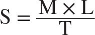
The Spreadability is calculated by following formula:
Where:
S=Spreadability (g·cm/s).
M=Weight applied (g).
L=Length (diameter) of the spread formulation (cm).
T=Time taken to spread the formulation (s).
A spreadability value of 6.88 g·cm/s is ideal because it ensures the gel spreads easily on the skin without being too runny or too thick, providing optimal application and retention.
Particle Size and Zeta Potential Determination
The particle size of the ethosomal formulation was found to be 370.8 nm with a Polydispersity Index (PDI) of 0.477, indicating nanoscale vesicles with uniform size distribution. This size range ensures good physical stability and reduces the risk of aggregation. It is ideal for topical applications, promoting efficient drug diffusion across the skin surface.
The zeta potential of the ethosomal formulation was found to be -18.6 mV, indicating a moderately negative surface charge. While lower than highly charged systems, this value still suggests reasonable electrostatic repulsion between vesicles, helping to reduce the risk of aggregation. The moderate stability is sufficient to maintain dispersion and prevent rapid coalescence, supporting the structural integrity of ethosomes. This level of stability is beneficial for short to moderate-term storage and helps preserve drug encapsulation efficiency. Hence, the analysis suggests that the prepared ethosomes are adequately stable for topical application.
In vitro release studies
The ethosomal gel was subjected to in vitro permeation studies to evaluate the release of the active ingredient at pH 5.5 over a period of 3 hr. The drug release study was conducted using a Franz diffusion cell maintained at 37ºC with a stirring speed of 350 rpm. The results of the in vitro permeation study of the ethosomal gel are presented in Figure 3. The in vitro drug release profile of the developed ethosomal gel formulation was compared with a plain Voriconazole gel over a period of 270 min. The graph clearly demonstrates a distinct difference in the drug release behavior of the two formulations. The plain Voriconazole gel exhibited a rapid and almost complete drug release, reaching over 90% release within 150 min, indicating a burst release pattern contrast, the ethosomal gel showed a slower, more controlled, and sustained release of the drug. The release was gradual, reaching 88.89% at 270 min, which suggests that the ethosomal vesicles effectively controlled the drug diffusion over time.
Antifungal Studies by Agar Well Diffusion Method
The antifungal activity of the developed ethosomal gel formulations was evaluated using the agar well diffusion method against Candida albicans. All samples including the standard Voriconazole solution and two ethosomal gel formulations (EG1 and EG2)-were tested at a concentration of 15 µg/mL. The results demonstrated that both ethosomal gel formulations exhibited significant antifungal activity, with zone of inhibition values comparable to that of the standard drug.
Although the standard Voriconazole solution exhibited a slightly larger zone of inhibition (27 mm±2 mm), the ethosomal gel formulations also showed significant antifungal activity with zones of inhibition of 24 mm±1 mm (EG1) and 22 mm±2 mm (EG2). This demonstrates that the ethosomal gel not only effectively incorporates the drug but also enhances its penetration and sustained release, resulting in comparable or even superior antifungal efficacy over time.
Antifungal study in Broth medium
Voriconazole solution (standard), Voriconazole-loaded ethosomes, and Voriconazole-loaded ethosomal gel were evaluated for antifungal activity against Candida albicans. Sabouraud Dextrose Broth (SDB) was poured into test tubes and sterilized by autoclaving at 121ºC for 20 min. After cooling, a fresh fungal inoculum of Candida albicans was added aseptically under laminar airflow. Each test formulation was then introduced into separate tubes. The tubes were incubated at 35ºC, and turbidity was observed as an indicator of fungal growth inhibition.
No turbidity was observed in any test tube, indicating absence of fungal growth.
Irritancy Studies
Irritancy studies were conducted to evaluate the dermal safety of the formulations. Hair was removed from the dorsal side of healthy rats using a commercially available depilatory cream 24 hr prior to sample application. The test, control, and marketed formulations were applied topically, and animals were observed for signs of erythema and edema at 24, 48, and 72 hr following the Draize scoring method.
The formula to calculate Primary Irritation Index:

Irritancy studies were performed on Wistar rats after hair removal using a depilatory cream on the dorsal surface. The animals were divided into three groups: Group I received the standard marketed preparation, Group II was treated with the ethosomal gel, and Group III received the plain gel. As per OECD guidelines, the formulations were applied and observed for signs of erythema and edema over 72 hr. Group III exhibited slight redness indicating mild irritation, while Group I and II showed no visible signs of dermal reaction (Table 4 and Figure 4). Mild erythema observed in the plain gel group may be attributed to the absence of phospholipid vesicles and ethanol-mediated skin conditioning effects, or slight irritation associated with excipients present in the gel base. On statistical analysis using ANOVA the p-value obtained on comparing standard and test group was found to be 0.804 and the p-value obtained on comparing test and control group was found to be 0.031.
| Formulation | Time (hr) | Mean Erythema Score | Mean Edema Score | Total Mean Score | PII | Draize Category |
|---|---|---|---|---|---|---|
| Marketed preparation (Darkatin) Standard | 24 | 0.33 | 0.17 | 0.50 | 0.50 | No Irritation |
| 48 | 0.00 | 0.00 | 0.00 | |||
| 72 | 0.00 | 0.00 | 0.00 | |||
| Ethosomal Gel (Test Formulation) | 0.00 | 0.17 | 0.17 | 0.34 | 0.34 | No Irritation |
| 48 | 0.00 | 0.00 | 0.00 | |||
| 72 | 0.00 | 0.00 | 0.00 | |||
| Plain Gel (Placebo Control Group) | 24 | 0.67 | 0.33 | 1.00 | 1.00 | Slightly irritant |
| 48 | 0.33 | 0.33 | 0.66 | |||
| 72 | 0.33 | 0.17 | 0.50 |
Stability Studies
The stability profile of Voriconazole was studied over a period of three months. It was observed that the drug remained stable throughout this period without any significant change in drug entrapment efficiency. The stability profile of the Voriconazole formulations is presented in Table 5.
| Time | Appearance | pH | % Entrapment Efficiency |
|---|---|---|---|
| Initial | Smooth, Homogeneous | 5.78 | 72.33%±1.38% |
| After 1 month | Smooth, Homogeneous | 5.80 | 72.02% + 2.31% |
| After 2 months | Smooth, Homogeneous | 5.75 | 71.4% + 1.67% |
| After 3 months | Smooth, Homogeneous | 5.80 | 70.2% + 1.15% |
The entrapment efficiency of the Voriconazole formulation showed only a slight decrease over three months, indicating good stability. Even after three months, the formulation retained 70.2% entrapment efficiency, demonstrating minimal drug loss and satisfactory performance over time.
DISCUSSION
Voriconazole, a BCS Class II drug with poor aqueous solubility, was successfully incorporated into ethosomal vesicles to enhance its topical delivery for the treatment of cutaneous candidiasis. The present study aimed to develop and evaluate a topical ethosomal gel formulation of Voriconazole for the effective treatment of cutaneous candidiasis. Ethosomes were selected as the vesicular drug carrier system to enhance the drug's retention and provide sustained diffusion from the topical formulation. Seven formulations were prepared using varying ratios of lecithin, cholesterol, ethanol, and aqueous phase components. Among these, Batch F7 was identified as the optimized formulation based on its favorable evaluation parameters.
The ethosomal suspension of Batch F7 exhibited a particle size of 370.8 nm, which falls within the nanometric range and is ideal for achieving uniform drug dispersion and smooth texture in topical formulations. The zeta potential was found to be -18.6 mV, indicating acceptable stability of the vesicles due to repulsive electrostatic forces that prevent aggregation. Although absolute zeta potential values greater than ±30 mV are generally associated with highly stable colloidal systems, the observed value of -18.6 mV indicates moderate electrostatic stabilization. The absence of significant aggregation during storage and the satisfactory stability profile suggest that steric stabilization provided by phospholipids and surfactant also contributed to formulation stability. The entrapment efficiency was high at 72.33%, confirming efficient loading of Voriconazole within the vesicular system. Additionally, the percentage yield of the formulation was 83.49%, reflecting efficient processing and minimal formulation loss. The particle size obtained in the present study (370.8 nm) was comparable to previously reported antifungal ethosomal formulations (200-500 nm). Similarly, entrapment efficiency of 72.33% falls within the range reported for phospholipid-based ethosomal systems.
Owing to these satisfactory parameters, the ethosomal suspension of F7 was successfully incorporated into a Carbopol 934P gel base to develop a semisolid topical formulation. The gel was evaluated for essential physical and chemical parameters and was found to possess a suitable pH of 5.78, which is skin-friendly and non-irritant. The viscosity was measured at different rpm. The viscosity of the gel was evaluated at different spindle speeds (RPMs) using a Brookfield viscometer, and the results indicated that the gel exhibited pseudoplastic behavior (shear-thinning), which is beneficial for topical application. This type of flow behavior ensures the gel spreads easily upon application and retains its structure afterward. The spreadability of the gel was found to be 6.88 g·cm/sec, suggesting that the gel has moderate to good spreading ability. This is important for ensuring the uniform distribution of the drug over the skin surface without requiring excessive rubbing, thus enhancing patient compliance.
In vitro diffusion studies using a Franz diffusion cell demonstrated that the ethosomal gel formulation provided a sustained drug release of 88.89% over 270 min, which supports prolonged therapeutic action and reduced dosing frequency. The release kinetics study revealed that the plain voriconazole gel followed first-order kinetics (R²=0.9845), indicating concentration-dependent drug release. In contrast, the voriconazole-loaded ethosomal gel showed the highest fit to the Korsmeyer-Peppas model (R²=0.99997) with a release exponent n=1.104, suggesting a Super Case-II transport mechanism involving polymer relaxation and swelling-controlled drug release. The sustained release profile of the ethosomal gel demonstrates the ability of the vesicular system to modulate drug release compared with the conventional gel formulation.
Antifungal activity studies against Candida albicans showed a significant zone of inhibition, confirming that the drug remained active after incorporation into the gel. In vivo skin irritancy testing using Wistar rats further supported the safety of the formulation, with no signs of erythema or edema observed and a Primary Irritation Index (PII) of 0.34, classifying it as non-irritant. Statistical evaluation shows the significant change in the PII of test and control group.
CONCLUSION
The present research focused on the design and development of a topical ethosomal gel of Voriconazole aimed at improving the therapeutic efficacy and skin retention of the drug in the treatment of cutaneous candidiasis. Voriconazole, being a BCS Class II drug, presents challenges in formulation due to its poor aqueous solubility. To address this, ethosomes were chosen as an advanced lipid-based delivery system, capable of enhancing the permeation and deposition of the drug through the stratum corneum into deeper layers of the skin.
The ethosomal gel was successfully formulated using phospholipids, cholesterol, ethanol, and surfactants, and was incorporated into a Carbopol 934 gel base. The optimized formulation showed satisfactory physicochemical characteristics and was further evaluated for in vitro drug release, antifungal activity, skin irritation, and stability. The ethosomal gel displayed sustained drug release, effective antifungal action, non-irritant behavior on animal skin, and maintained physical and chemical stability under storage conditions.
The findings suggest that ethosomal gels can be a reliable and patient-compliant alternative to conventional topical formulations. This formulation can help minimize dosing frequency, improve local drug concentration, and avoid systemic side effects, thereby offering an effective solution for superficial fungal infections like cutaneous candidiasis. Overall, the ethosomal system offers a viable platform for enhancing the performance of poorly soluble antifungal drugs and can be considered a step forward in topical drug delivery technology.
